Aneurysms
Aneurysms
The word is from Greek:" aneurysma" mean, "a widening "a dilation", from , aneurynein, "to dilate
Most aneurysms occur in the aorta, but they can also occur in other arteries in the brain, heart, intestines, neck, spleen, back of the knees and thighs, or elsewhere in the body. If an aneurysm in the brain bursts, it can cause a stroke.Aneurysm is the second most frequent disease of the aorta after atherosclerosis.
Definition-
- An aneurysm is an outward bulging, likened to a bubble or balloon, caused by a localized, abnormal, weak spot on a blood vessel wall. Aneurysms may be a result of a hereditary condition or an acquired disease.
- According to American heart association an aneurysm occurs when part of an artery wall weakness, allowing it to widen abnormally or balloon out.
- Localized abnormal dilation of blood vessel or heart due to blood vessel wall stress.
- Weak spot on a blood vessel wall that causes an outward bulging, likened to a bubble or balloon
Classification of Aneurysm
A. According to Size or Morphology -
- Fusiform Aneurysm- It is a diffuse dilation that involves the entire circumstances of the arterial segment. i.e., whole artery.
- Saccular Aneurysm- It is a distention of a vessel projecting from one side. It is a distinct, localized out pouching of the arterial wall.
- Dissecting Aneurysm- Haemorrhage or intramural hematoma, separating the layers of an arterial wall. It commonly involves Arch of aorta.

B. According to cause-
- True Aneurysm- Involve all three layers of wall of artery. It is a result of the slow weakening of the arterial wall caused by long term diseases such as hypertension, atherosclerosis, etc.
- False Aneurysm- Pseudoaneurysm is caused by traumatic break in the arterial wall. mostly outer later of artery involved.
C. Based on Location- Types of aneurysm include thoracic and abdominal aortic aneurysms, cerebral aneurysms and peripheral aneurysms.
1. Aortic aneurysm- Most aneurysms occur in the aorta. The aorta is the main artery that carries blood from the heart to the rest of the body. The aorta comes out from the left ventricle (main pumping area) of the heart and travels through the chest and abdomen.
- Thoracic Aortic Aneurysm- An aortic aneurysm that occurs in the part of aorta running through the thorax (chest) is called a thoracic aortic aneurysm (TAA). One in four aortic aneurysms is a TAA. The most common feature of the Thoracic aortic aneurysm is severe pain, constant, in supine posture.
- Abdominal Aortic Aneurysm- An aortic aneurysm that occurs in the part of the aorta running through the abdomen is called an abdominal aortic aneurysm. Three out of four aortic aneurysms are AAAs. Abdominal aortic aneurysm most commonly occurs at infra-renal part of aorta.
2. Cerebral Aneurysm- Aneurysms that occur in an artery in the brain are called cerebral aneurysms. They are sometimes called berry aneurysms because they are often the size of a small berry. Most cerebral aneurysms produce no symptoms until they become large, begin to leak blood, or rupture. A ruptured cerebral aneurysm causes a stroke.
3. Peripheral Aneurysm -Aneurysms that occur in arteries other than the aorta (and not in the brain) are called peripheral aneurysms. Common locations for peripheral aneurysms include the popliteal artery that runs down the back the thigh behind the knee; the femoral artery, which is the main artery in the groin; and the carotid artery, which is the main artery in the neck.
Etiology
- Atherosclerosis
- Heredity
- Infection
- Trauma
- Immunologic conditions
- Hypertension
- Arteriosclerosis
- Local infections
- Syphilis
- Trauma (pseudoaneurysm)
- Congenital disorders include primary connective tissue disorders.
Clinical Manifestations
- Aneurysm of the thora or abdominal aorta it includes: marked pulse and blood pressure differences
- Chest pain
- Dyspnea
- Cough
- Hoarseness of voice
- Edema of chest wall
- Cyanosis
- Dysphagia
Abdominal aneurysm includes symptoms such as-
- Abdominal and lower back pain
- Distal variability of blood pressure
- Hypertension
- Feeling of abdominal pulsating sound on auscultation
Cerebral Aneurysm-
- Mild to severe headaches
- Vision impairment
- Pain above and behind the eye
- Nausea and vomiting
- Drooping eyelid
- Dilated pupil
- Paralysis or numbness of one side of the face
- Sensitivity to light
- Stiffness or Pain in neck
- Seizures
Pathophysiology
Increases wall stress➡️Weakening of vessel wall➡️Outpouching of vessel wall➡️ abnormal dilatation of vessel wall➡️Aneurysm
Diagnostic Evaluation-
- An aneurysm may be diagnosed by chance during a routine physical examination
- Chest/Abdominal X-ray
- Ultrasound
- Echocardiography
- Computed tomography
- Magnetic resonance imaging (MRI)
- Angiography
- Aortogram
Management
The goals of management may include:
- Preventing the aneurysm from growing
- Preventing or reversing damage to other body structures
- Preventing or treating a rupture or dissection
- Allowing the pt. to continue doing their normal daily activities
- Smoking Cessation- Single most important modifiable risk factor
- Exercise Therapy- Evidence suggests may benefit small aneurysms
- Beta Blockers- May decrease the rate of expansion.eg-Atenolol
- ACE (Angiotensin-converting enzyme)inhibitors- Implicated in less aneurysm rupture.
- Doxycycline- Against chlamydia species
- Statins- associated with reduced aneurysm expansion rates
Surgical Management
Surgery may be recommended if an aneurysm is large and likely to rupture. Enlarging thoracic aneurysms should be considered for surgery. Two main types of surgery to repair aortic aneurysms are Open abdominal or Open chest repair and Endovascular repair.
1. Open Repair-
- The traditional and most common type of surgery for aortic aneurysms is open abdominal or open chest repair. It involves a major incision in the abdomen or chest. General anaesthesia is needed with this procedure. The aneurysm is removed and the section of aorta is replaced with an artificial graft made of material such as Dacron or Teflon.
- The surgery takes three to six hours, and the patient remains in the hospital for five to eight days. It often takes a month to recover from open abdominal or open chest surgery and return to full activity. Open abdominal and chest surgeries have been performed for 50 years. More than 90% of patients make a full recovery.
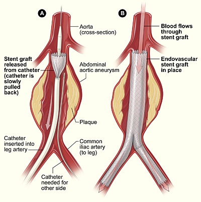
2. Endovascular Repair-
- In this, the aneurysm is not removed, but a graft is inserted into the aorta to strengthen it. This type of surgery is performed through catheters inserted into the arteries; it does not require surgically opening the chest or abdomen.
