The cardiovascular system
Heart
Heart is a hollow muscular organ that pumps blood throughout the circulatory system. It is situated in between two lungs in the mediastinum. It is made up of four chambers, two atria and two ventricles.
The apex is about 9 cm to the left of the midline at the level of the 5th intercostal space, i.e. a little below the nipple and slightly nearer the midline. The base extends to the level of the 2nd rib.
- Shape-Cone with Broad base and Narrow Apex
- Size (close fist)-Length-12cm
- Width-9cm
- Thickness -6cm
- Weight- Adults Male-300gram
- Adults female 250gram
Organs associated with the heart-
- Inferiorly - The apex rests on the central tendon of the diaphragm
- Superiorly - The great blood vessels, i.e. the aorta, superior vena cava, pulmonary artery and pulmonary veins
- Posteriorly - The oesophagus, trachea, left and right bronchus, descending aorta, inferior vena cava and thoracic vertebrae
- Laterally -The lungs — the left lung overlaps the left side of the heart
- Anteriorly - The sternum, ribs and intercostal muscles

Right side of the Heart-
Right side of the heart has two chambers,
- Right atrium
- Right ventricle
Right atrium is a thin walled thickness is 2-3 mm and low-pressure chamber. It has got the pacemaker known as sinoatrial node that produces cardiac impulses and atrioventricular node that conducts the impulses to the ventricles.
Right atrium receives venous (deoxygenated) blood via two large veins-
- Superior vena cava that returns venous blood from the head, neck and upper limbs
- Inferior vena cava that returns venous blood from lower parts of the body
Right atrium communicates with right ventricle through Tricuspid valve. Wall of right ventricle is thick. Venous blood from the right atrium enters the right ventricle through this valve. From the right ventricle, pulmonary artery arises. It carries the venous blood from right ventricle to lungs. In the lungs, the deoxygenated blood is oxygenated.
Left side of the Heart-
Left side of the heart has two chambers-
- left atrium
- left ventricle
- Left atrium is a thin walled and low-pressure chamber. It receives oxygenated blood from the lungs through pulmonary veins. This is the only exception in the body, where an artery carries venous blood and vein carries the arterial blood. Blood from left atrium enters the left ventricle through Mitral valve (bicuspid valve). Wall of the left ventricle is very thick walled thickness is 10-15 mm.
- Left ventricle pumps the arterial blood to different parts of the body through systemic aorta. and left ventricles are separated from one another by interventricular septum. The upper part of this septum is a membranous structure, whereas the lower part of it is muscular in nature.
Layers of the Heart-
Heart is made up of three layers of tissues-
- Outer pericardium
- Middle myocardium
- Inner endocardium.

A. Outer pericardium-
Pericardium is the outer covering of the heart. It is made up of two layers-
- Outer parietal pericardium
- Inner visceral pericardium.
The space between the two layers is called pericardial cavity or pericardial space and it contains a thin film of fluid.
- Outer Parietal Pericardium
Parietal pericardium forms a strong protective sac for the heart. It helps also to anchor the heart within the mediastinum.
Parietal pericardium is made up two layers-
- Outer fibrous layer
- Inner serous layer.
2. Inner Visceral Pericardium
Inner visceral pericardium lines the surface of myocardium. It is made up of flattened epithelial cells. This layer is also known as epicardium.
B. Middle myocardium-
Myocardium is the middle layer of wall of the heart and it is formed by cardiac muscle fibers or cardiac myocytes. Myocardium forms the bulk of the heart and it is responsible for pumping action of the heart.
Myocardium has three types of muscle fibers-
- Muscle fibers which form contractile unit of heart - Cardiac muscle fiber is bound by sarcolemma. It has a centrally placed nucleus. Myofibrils are embedded in the sarcoplasm. Sarcomere of the cardiac muscle has all the contractile proteins, namely actin, myosin, troponin and tropomyosin. Sarcotubular system in cardiac muscle is similar to that of skeletal muscle.
- Muscle fibers which form pacemaker- Pacemaker is structure in the heart that generates the impulses for heart beat. It is formed by pacemaker cells called P cells. Sinoatrial (SA) node forms the pacemaker in human heart.
- Muscle fibers which form conductive system- Impulses from SA node is transmitted to the atria directly. However, the impulses are transmitted to ventricles through various components of conducting system,
C. Endocardium-
This forms the lining of the myocardium and the heart valves. It is a thin, smooth, glistening membrane which permits smooth flow of blood inside the heart. It consists of single layer of squamous epithelium tissue.
Valves of the Heart-
There are four valves in human heart. Two valves are in between atria and the ventricles called atrioventricular valves. Other two are the semilunar valves, placed at the opening of blood vessels arising from ventricles, namely systemic aorta and pulmonary artery.

1. Atrioventricular Valves
Left atrioventricular valve is otherwise known as mitral valve or bicuspid valve. It is formed by two valvular cusps or flaps. Right atrioventricular valve is known as tricuspid valve and it is formed by three cusps.
2. Semilunar Valves
Semilunar valves are present at the openings of systemic aorta and pulmonary artery and are known as aortic valve and pulmonary valve respectively. Because of the half-moon shape, these two valves are called semilunar valves. Semilunar valves are made up of three flaps.
Blood supply to the heart
1. Arterial supply-
The heart is supplied with arterial blood by the right and left coronary arteries which branch from the aorta immediately distal to the aortic valve.The coronary arteries receive about 5% of the blood pumped from the heart, although the heart comprises a small proportion of body weight. This large blood supply, especially to the left ventricle, highlights the importance of the heart to body function.
2. Venous drainage
Most of the venous blood is collected into several small veins that join to form the coronary sinus which opens into the right atrium. The remainder passes directly into the heart chambers through little venous channels.
Conducting system of the Heart
The heart has an intrinsic system whereby the cardiac muscle is automatically stimulated to contract without the need for a nerve supply from the brain. However, the intrinsic system can be stimulated or depressed by nerve impulses initiated in the brain and by circulating chemicals including hormones. There are small groups of specialised neuromuscular cells in the myocardium which initiate and conduct impulses causing coordinated and synchronised contraction of the heart muscle.
A. Sinoatrial node (SA node)
This small mass of specialised cells is in the wall of the right atrium near the opening of the superior vena cava. The SA node is the 'pace-maker' of the heart because it normally initiates impulses more rapidly than other groups of neuromuscular cells.
WHY SA NODE LEADS THE HEART?
| TISSUE | RATE OF IMPULSE GENERATION |
| SA NODE | 70-80/MIN |
| AV NODE | 40 – 60/MIN |
| BUNDLE OF HIS | 40/MIN |
| PURKINJE SYSTEM | 24/MIN |
B. Atrioventricular node (AV node)
This small mass of neuromuscular tissue is situated in the wall of the atrial septum near the atrioventricular valves. Normally the AV node is stimulated by impulses that sweep over the atrial myocardium. However, it too is capable of initiating impulses that cause contraction but at a slower rate than the SA node.
C. Atrioventricular bundle (AV bundle or bundle of His)
This is a mass of specialised fibres that originate from the AV node. The AV bundle crosses the fibrous ring that separates atria and ventricles then, at the upper end of the ventricular septum, it divides into right and left bundle branches. Within the ventricular myocardium the branches break up into fine fibres, called the Purkinje fibres. The AV bundle, bundle branches and Purkinje fibres convey electrical impulses from the AV node to the apex of the myocardium where the wave of ventricular contraction begins, then sweeps upwards and outwards, pumping blood into the pulmonary artery and the aorta.
Cardiac cycle-
Cardiac cycle is defined as the succession of (sequence of) coordinated events taking place in the heart during each beat. Each heartbeat consists of two major periods called systole and diastole.
- During systole, heart contracts and pumps the blood through arteries.
- During diastole, heart relaxes and blood is filled in the heart.
Events of cardiac cycle are classified into two
- Atrial events
- Ventricular events.
Duration of cardiac Cycle-
When the heart beats at a normal rate of 72/minute, duration of each cardiac cycle is about 0.8 second.
Atrial events are divided into two divisions-
- Atrial systole = 0.11 (0.1) sec
- Atrial diastole = 0.69 (0.7) sec.
Ventricular events are divided into two divisions-
- Ventricular systole = 0.27 (0.3) sec
- Ventricular diastole = 0.53 (0.5) sec.
Heart sounds
The individual is not usually conscious of his heartbeat, but if the ear or the diaphragm of a stethoscope is placed on the chest wall a little below the left nipple and slightly nearer the midline the heartbeat can be heard. Two sounds, separated by a short pause, can be clearly distinguished. They are described in words as 'lub dup'. The first sound, 'lub', is fairly loud and is due to the closure of the atrioventricular valves. This corresponds with ventricular systole. The second sound, 'dup', is softer and is due to the closure of the aortic and pulmonary valves.
| LUB | DUB |
| Due to closer of A.V. valves | Due to closer of Semilunar valves |
| Systolic sound | Diastolic sound |
| Longer duration 0.14 second | Shorter duration 0.11 second |
| Louder | Less loud |
| Durng ventricular Systole | Durng ventricular Diastole |
Cardiac Output-
Cardiac output is the amount of blood pumped from each ventricle. Usually, it refers to left ventricular output through aorta.
Usually, cardiac output is expressed in three ways-
- Stroke volume
- Minute volume
- Cardiac index.
A.Stroke volume- Stroke volume is the amount of blood pumped out by each ventricle during each beat. Normal value: 70 mL (60 to 80 mL) when the heart rate is normal (72/minute).
B. Minute volume- Minute volume is the amount of blood pumped out by each ventricle in one minute. It is the product of stroke volume and heart rate-
- Minute volume = Stroke volume × Heart rate
- Normal value- 5 L/ventricle/minute.
C. Cardiac Index-Cardiac index is the minute volume expressed in relation to square meter of body surface area. It is defined as the amount of blood pumped out per ventricle/minute/ square meter of the body surface area. Normal value: 2.8 ± 0.3 L/square meter of body surface area/minute (in an adult with average body surface area of 1.734 square meter and normal minute volume of 5 L/minute).
Circulation
Double circulation
In human beings, the blood goes through the heart twice during each cycle i.e. the blood passes through the human heart two times to supply once to the whole body. So, it is called Double Circulation of blood.
The double circulation of blood includes-
- Systematic Circulation
- Pulmonary Circulation
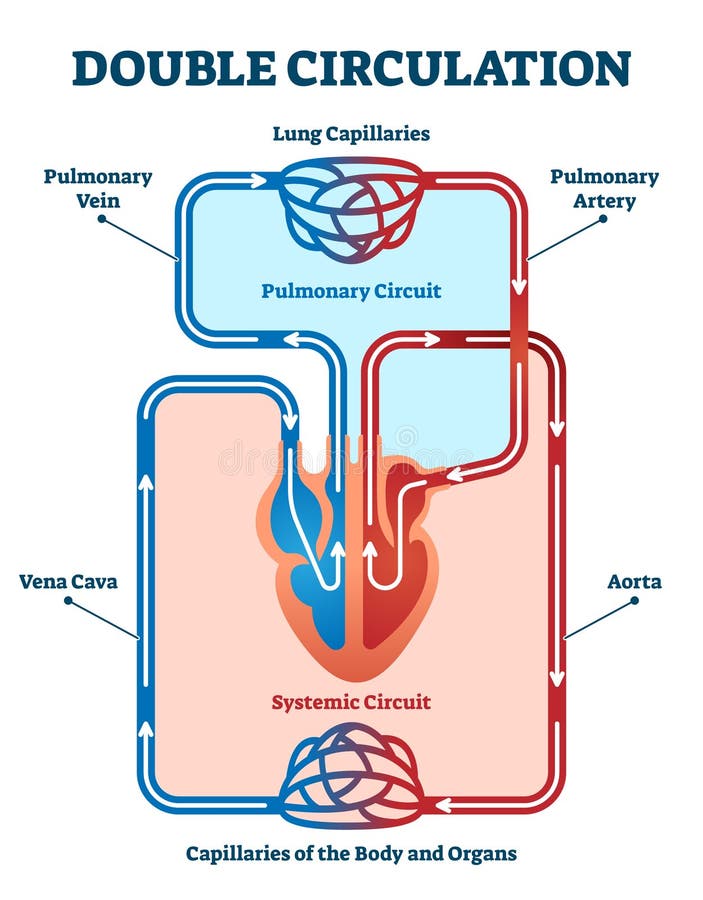
1. Systematic Circulation- (Body>Heart>Body)
- The oxygenated blood that returns to left atrium, moves to left ventricle and when the left ventricles contracts, the blood is forced into the aorta and circulate into the whole body, via systemic arteries and supply oxygen to all tissues. Finally, the blood returns back to the right atrium via the superior and inferior vena cava as deoxygenated blood and moves into right atrium. This circulatory pattern from left ventricle to right atrium is called the Systemic or Peripheral or Greater circulation.
2.Pulmonary Circulation- (Heart>Lungs >Heart)
- The deoxygenated blood is pumped by the right ventricle into the lungs for oxygenation. The oxygenated blood is brought back to left atrium (auricle) of the human heart. From left atrium, the oxygenated blood is pushed into the left ventricle.
- The pulmonary circulation is situated in series with the heart and receives all the blood ejected from heart. The pulmonary circulation brings blood to the alveoli of lungs so that gaseous exchange can occur and the series arrangement ensure the oxygenation of the total amount of blood ejected from heart.