Nervous system
Nervous system
Nervous system controls all activity of the body it is quicker than the other control system in the body like– endocrine system.
Primarily nervous system divided into 2 parts
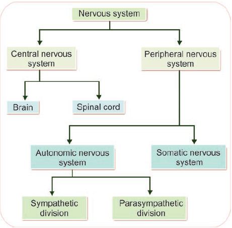
- C.N.S – central nervous system
- P.N.S – peripheral nervous system
1. C.N.S:– C.N.S includes brain and spinal card it is formed by neurons (nerve cell) and supporting cell called neuroglia.
- Structure of brain and spinal cord are arranged in the layers namely is gray matter and white matter.
- Gray matter formed- nerve cell body (soma) + dendrites + unmylinated axon+ capillary.
- White matter- made by only mylinated axon,
- Brain is situated in the skull. It is continued as spinal card in the vertebral canal through foramen magnum of the skull bone.
Largest holl of skull- form magnum.
Brain and spinal cord are surround by 3-layers of meninges called.
- Outer- dura mater
- Middle- arachnoid matter
- Inner- pia matter
Space b/w arachnoid matter and pia matter is called subarachnoid spaces. This space fill with fluid is known as cerebrospinal fluid.
2. P.N.S- peripheral nervous system- it consists of cranial nerve (12 pair) arising from brain and spinal nerve (31 pair) arises from the spinal cord. It again divided in 2-types
A. Somatic nervous system.
B. Autonomic nervous system.
A. Somatic nervous systems- it includes nerve supply and control movements of body.
B. A.N.S- it regulate function of visceral organ and vegetative function other name- vegetative or involuntary nervous systems. Again divided in two –
- Sympathetic
- parasympathetic
Neuron: – Neuron is defined as the structural and functional unit of the nervous system. Ti tis otherwise k/w nerve cell neuron is like any other cell in the body having nucleus and all the organelles in the cytoplasm.
- Basic functional cell of nervous system
- Transmits impulses (up to 250 mph)
However it is different from other cell by 2- ways
1. Neuron has branches of process called axon and dendrites.
2. Neuron does not have centrosome so it cannot undergo division (not regenerate)
Classification of neuron: - Neurons are classified by 3- methods:-
- Depending upon the number of poles
- Depending upon the function
- Depending upon the length of the axon
1. Depending upon the number of poles:-
A. Unipolar neurons:- have only one pole. From single arise. This type cell present only in embryonic stage in human beings.
B. Bipolar neurons – neurons with 2-poles. Axon arises from one pole and dendrites arise from another pole.
C. Multipolar neurons- the neurons which have many poles. One of the poles gives rise to the axone and all other pole give rise dendrities.
2. Depending up on the funtions- 2 types
A. Motor neuron
B. Sensory neuron
A. Motor neuron:-
- Motor neuron is also k/w efferent nerve cell/ efferent neuron.
- These neuron calley motor impulse from C.N.S to peripheral effector organ. Like- muscles, gonodes.
- Motor neurons has long axons and short dendrites.
B. sensory neuron:-
- sensory neurons are also called afferent nerve cell/ afferent neuron
- these neurons carry the sensory impulse from periphery to C.N.S
- sensory neuron has short axon and long dendrites
3. depending upon the length of axon neuron are 2-type:-
- Golgi type- I neurons.
- Golgi type- II neuron.
a. Golgi type- I neuron have long axons. The cell body of these neuron is in C.N.S and axons reach the remote peripheral organs
b. Golgi type- II – neurons of this type have short axons. These present in cerebral cortex and spinal cord.
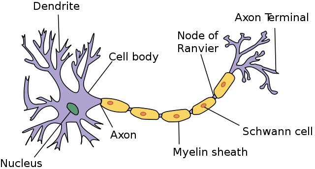
Structure of neuron:- neuron is made up of 3 parts
1. Nerve cell body
2. Dendrite
3. Axon
Dendrites and axon form the process of neuron dendrites are short process and axon are long process.
Most- dendrites and axons are usually called nerve fibers
Mylinated nerve fibers – nerve cover which sheath is called mylinated nerve fiber.
Myline sheath- myline sheath is not a continuous sheath. It is absent at regular inter well. The area where myline sheath absent is called node of Ranvier myeline sheath is responsible for white colour of nerve fibers. It is formed by schwann cell.
Mylinesheath is work as insulator (means – not current pass)
Classification of nerve fiber
1. Depending up on structure: –
a. Mylinated nerve fibers- these covered by myelin sheath.
b. Hon myelinated nerve fibers – they do not have myelin sheath.
2. Depending up on distribution: –
a. Somatic nerve fiber- which fibers supply skeletal muscle of body.
b. Visceral/autonomic nerve fibers- supply internal organ of body.
3. Depending up on origin:-
a. Cranial nerves – which nerve fibers arising from brain.
b. Spinal nerve – whicharising from spinal cord.
4. Depending up on function:- ( efferent )
a. Motor/efferent nerve fibers- from C.N.S to different body part.
b. Sensory nerve fibers – these carry sensory impulse from different body
part to C.N.S.
5. Depending up on secretion of neurotrons metter:-
a. Adrenergic nerve fibers – which secrete norade narrow
b. Cholinergic – which secrete acetylecholine.
Neuroglia
Neuroglia is the supporting cell of the nervous system. Neuroglia cells are non-excitable and do not transmit. Have impulse so these cells called nonneural cell.
Classification of neuroglia cells- neuroglia cells are distributed in C.N.S as well as P.N.S acc. To this these are two types.-
1. Central neuroglia cell
2. Peripheral neuroglia
1. C.N. cell- are 3 types
a. Astrocytes
b. microglia
c. oligodorodrocytes
a. Astrocytes – astrocytes are star shape cell present in all part of brain. 2- types of astrocytes
- Fibrous astrocytes – occapise mainly in white matter process of these cells cover the neuron synapses, and form B.B. Barrier. By sending process to blood vessels of brain. Particulary the capillaries.
- Protoplasmic astrocytes – protoplasmic astrocytes are present mainly in gray matter.
b. Microglia-
- Microglias are the smallest neuroglial cells.
- These cells derived from monocytes and enter into nervous system from blood.
- These phagocytic cell migrate to the site of injection or injury.
- Often called macrophages of C.N.S.
c. Oligodendrocytes – these cells produce myelin sheath around the nerve fibers in C.N.S.
2. Peripheral neuroglial cell- 2 types
a. Schwann cell
b. satellite cell
a. Schwann cell – provide myelination around the nerve fiber in PNS.
b. Satellite cell- presents on exterior surface of PNS neurons.
Function- provide physical support to PNS neurons
Receptors – receptors are sensory (afferent) nerve endings that give responses to the stimulas. When stimulated receptors produce a series of impulses, which are transmitted through the afferent nerves.
Classification of receptorsReceptors are classified into 2 types
1. Extereceptors
2. Introceptors
1. Exteroceptors – are the receptors which give response to stimli arising from outside the body. The 3-types of receptor
A. Cutaneous/ mechano receptors- these receptors situated in skin liketouch, pressure, pain
B. Chemoreceptors – receptors which give response to chemical stimule.
C. Telereceptors – these give response to stimuli arising away from the body. Like- vision, hearing.
2. Interoceptors- these receptors give response to stimuli arising within the body. These receptor 2-types
a. Visceroceptors – situated in the viscera. Like- stretch receptors- heart Baroreceptors- blood vessels.
b. Proprioceptors-this receptor give response to change in position of different parts of body.
Synapse: -
Junction between two neurons is called synapse:- it is not the anatomical continuation. But is only a physiological continuity between 2-neuron.
Classification of synapse
1. Anatomical classification
2. Physiological classification
1. Anatomical classification –
- Axosomatic synapse - axmol one neuron end on some (cell body) of another neuron.
- Axodendrite synapse – axon of one neuron end on dendrite of another neuron.
- Axoaxonic synapse – axon of one neuron end on axon of another neuron.
2. Functional classification –
a. Electrical synapse
b. Chemical synapse
a. Electrical synapse- physiological continuity b/w presynaptic and post synaptic neurons is provided by Gap junction.
- So direct exchange of irons b/w two neuron.
- Important - ↓se synaptic delay.
- Commany found in nervous system, cardiac muscle fiber, smooth muscle fibers of intestine.
b. Chemical synapse-
- Mitrochndria – which help in synthesis of neurotransmitter
- Synaptic vesicle- which store the neurotransmetters.
- Post synaptic membrane contain receptors.
- Space b/w presynaptic and post synaptic neuron is called synaptic cleft.
- Synaptic cleft has cholinesterase which destroy ach.
Properties of synapse –
- One way conduction (Bell- magendie law)-Impulse are transmit only in one direction that is from presynaptic neuron to post synaptic.
- The synaptic delay - Due to Release of neurotransmitter (due to action potential). Movements of neurotransmitter from axon turinal to post synaptic membrain. Action of neurotransmitter on post synaptic membrain to open ions charnels.Synaptic delay is one of the cause for latent period of reflex activity.
- Fatigue- continue muscle activity faligue due to ↓se ach.
- Cause- Achs destroyed by acetylcholineesterage. Due to continuous action, new ach is not synthesized.
Parts of brain –
Brain consists 3 – major division
1. Prosencehalon
2. Mesencephalon
3. Rhombencephalon

1. Prosencephalon – also k/w fore brain. It is further divided into two parts.
- Telencephalon –which includes – cerebral hemispheres, basalganglia,hippocampus.
- Dienccphalons – thalamus, hypothalamus, subthalum, metathalamus,
2. Mesencepahalone- also called midbrain
3. Rhombencephalon – also called hindbrain subarea
a. Metencephalon- formed by pones and cerebellum
b. Myelmchalone – medulla oblongata.
Cerebral cortex– (cerebrum)
Introcuction- the cerebral cortex is also called pallidum. In human beings, it has an area of 2.2 sqm. This is the largest part of the brain situated in the anterior and middle cranial fossa.
- During embryonic development, the brain size increase rapidly, the gray matter of the cortex enlarge much faster than the deeper white matter. As a result the cortical region rolls and folds upon itsey. The folds are known as gyri.
- The deepest grooves between folds are known as fissures, the shallower (small) grooves between folds are termed sulci.
- The most prominent fissure, the longitudinal fissure separates the cerebrum into right and left cerebral hemispheres. The hemispheres the hemispheres are connected inter nally by the corpus callosum, a broad band of white matter.
Note- cerebral cortex consists of gray matter that surrounds the deeper white cortex. White the spinal cord outer part contain white matter that surround. Inner part gray matter.
Lobes of the cerebrum– each cerebral hemisphere card be further subdivided into 4-lobes. The lobes are named after the bones that cover them. Frontal, parital, temporal and occipital lobes.
- The central sulcus separates the frontal lobe (rolandic fissure) from the parietal lobe.
- The lateral sulcus separates the frontal lobe from (sylvian fissure) the temporal lobe.
- Parieto-occipital sulcus separates the parietal lobe from the occipital lobe.

1. Frontal lobe functional areas
a. Primary motor area-
4→area no. 4 for initiation of voluntary movements
4s→(suppressor area) → is suppresses the extra impulses produce by area 4 and prevent exaggeration of movements.
b. Premotor area – this has areas 6,8,44, 45
- Area no. 6→ it is concerned with coordination of movements initiated by area-4.
- Area no. 8 → it concerned with movements of eyeball.
- Area no 44, 45 (broca’s area)- this area responsible for speech.
2. Parital lobe functional area-
- Area no. 1, 2, 3- these are sensory area. These area receive nerve impulse for touch, proprioception (joint and muscle), pain itching, tickle, and temperature and is involved in the perception of these sensations.
- Area no. 43 (gustatory area)- this area receive impulses for taste.
3. Occipital lobe functional area-
- Area no. 17 (primary visual area) – this area responsible for visual perception.
- Area no. 18 (visual association area)- this is responsible for interpretation of visual perception
- Area no. 19 (occipital eye field) – this area responsible for movements of eye.
4. Temporal lobe functional areas-
- Area no. 41, 42 (primary auditory area)/ (Wernicke’s area)This area responsible for the hearing. This perceives auditory impulses.
- Area no. 22(auditopsychic area) – this area responsible for interpretation of auditory impulse perceive by wernicke’s area)
- Area no. 28 (olfactory area) this involved in olfactory perception. This responsible for smell.
Brainstem–
Brainstem is the part of brain formed by medulla oblongata, pons and midbrain. Brainstem contains ascending and descending tracts between brain and spinal cord. It also contains many centers for regulation of vital functionsin the body.
Midbrain –
Midbrain is the area of brain situated around the cerebral aqueduct between the cerebrum above and the pons below it consists of two parts:
a. Tectum-tectum is formed by two structures:-
- superior colliculus
- Inferior colliculus.
- superior colliculus – superior colliculus is a small structure and is an important center for reflexes
- Inferior colliculus – inferior colliculus is the center for auditory reflexes.
b Cerebral peduncles –
- Pedunculus consists of pyramidal tract fibers in the middle, temporopontinefibers laterally and frontopontine fibers medially.
Pons-
Pons forms a bridge between medulla and midbrain. It is situated in front of the cerebellum, below the midbrain and above the medulla oblongata.
Functions of pons
1. pons forms the pathway that connects cerebellum with cerebral cortex.
2. Pyramidal tracts pass through the pons
3. nuclel of 8th, 7th, 6th, and 5th cranial nerves are located in pons
4. pons contains the pneumotaxic and apneustic centers for regulation of respiration.
Medulla oblongata-
- Medulla oblongata is continuous with the superior part of spinal cord and it forms the inferior part of brain stem. The medulla begins at the foramen magnum and extend to the inferior border of pons.
- The medulla white matter contains all sensory (ascending) and motor (descending) tract that extend between the spinal cord and other part of brain. Some of white matter forms bulges on the anterior aspect of the medulla. The protrusions called the
- pyramids. Just superior to junction of medulla with the spinal cord 90% of axons in the left pyramid cross to right side and 90% off axon in right pyramid cross to left side. This crossing called the decussation of pyramid.
Function of medulla oblongata:-
1. Respiratory centers.
2. Vasomotor center: - vasomotor center controls blood pressure and heart rate.
3. Deglutition center – deglutition center regulates the pharyngea and esophageal stages of deglutition.
4. Vomiting center – vomiting center vomiting center induces vomiting during ittitation or inflammation of gastrointestinal (GI) tract.
5. Superior and inferior salivatory nucle – salivatory nuclei control the secretion of saliva.
6. Cranial nerve nuclei- nuclei of 12th, 11th, 10th, and some nuclei of 8th and 5th cranial nerves are located in the medulla oblongata.
Cerebellum–
The cerebellum is situated behind the pons and immediately below the posterior portion of the cerebrum occupying the posterior cranial fossa.
Parts of cerebellum-
Cerebellum consists of a narrow, worm-like central body called vermis and two lateral lobes, the right and left cerebellar hemispheres.
Vermis
- Vermis of cerebellum is formed by nine parts.
- Cerebellar hemispheres –Cerebellar hemispheres are the extended portions on either side of vermis.
SPINAL CORD ANATOMY
Protective Structures of spinal cord
Two types of connective tissue coverings—bony vertebrae and tough, connective tissue meninges—plus a cushion of cerebrospinal fluid (produced in the brain) surround and protect the delicate nervous tissue of the spinal cord.
Meninges
- The meninges are three connective tissue coverings that encircle the spinal cord and brain. The spinal meninges surround the spinal cord and are continuous with the cranial meninges, which encircle the brain.
- The most superficial of the three spinal meninges, the dura mater is composed of dense, irregular connective tissue.
- The spinal cord is also protected by a cushion of fat and connective tissue located in the epidural space, a space between the dura mater and the wall of the vertebral canal.
- The middle meninx is an avascular covering called the arachnoid mater. Between the
- dura mater and the arachnoid mater is a thin subdural space, which contains interstitial fluid.
- The innermost meninx is the pia mater a thin transparent connective tissue layer that adheres to the surface of the spinal cord and brain.Within the pia mater are many blood vessels that supply oxygen and nutrients to the spinal cord. Between the arachnoid mater and the pia mater is the subarachnoid space, which contains cerebrospinal fluid.
External Anatomy of the Spinal Cord
- The spinal cord, although roughly cylindrical, is flattened slightly anteriorly and posteriorly. In adults, it extends from the medulla oblongata, the inferior part of the brain, to the superior border of the second lumbar vertebra. In newborn infants, it extends to the third or fourth lumbar vertebra.
- During early childhood, both the spinal cord and the vertebral column grow longer as part of overall body growth. Elongation of the spinal cord stops around age 4 or 5, but growth of the vertebral column continues. Thus, the spinal cord does not extend the entire length of the adult vertebral column.
- The length of the adult spinal cord ranges from 42 to 45 cm. Its diameter is about 2 cm in the mid thoracic region, somewhat larger in the lower cervical and mid lumbar regions, and smallest at the inferior tip. The spinal cord terminates as a tapering, conical structure called the conus medullaris which ends at the level of the intervertebral disc between the first and second lumbarvertebrae in adults. Arising from the conus medullaris is the filum terminale, an extension of the pia mater that extends inferiorly and blends with the arachnoid mater and dura mater and anchors the spinal cord to the coccyx.
- The gray matter on each side of the spinal cord is subdivided into regions called horns. The posterior gray horns contain cell bodies and axons of interneurons as well as axons of incoming sensory neurons. The anterior gray horns contain somatic motor nuclei, which are clusters of cell bodies of somatic motor neurons that provide nerve impulses for contraction of skeletal muscles. Between the posterior and anterior gray horns are the lateral gray horns, which are present only in thoracic and upper lumbar segments of the spinal cord. The white matter of the spinal cord, like the gray matter, is organized into regions. The anterior and posterior gray horns divide the white matter on each side into three broad areas called columns:
1 anterior white columns
2 posterior white columns, and
3 lateral white columns.
spinal nerves
- spinal nerves are the paths of communication between the spinal cord and specific regions of the body. The spinal cord appears to be segmented because the 31 pairs of spinal nervese merge at regular intervals from intervertebral foramina. but, for convenience, the naming of spinal nerves is based on the segment in which There are 8pairs of cervical nerves 12 pairs of thoracic nerves (T1–T12), 5 pairs of lumbar nerves(L1–L5), 5 pairs of sacral nerves (S1–S5), and 1 pair of coccygea lnerves (Co1).Because the spinal cord is shorter than the vertebral column, nerves that arise from the lumbar, sacral, and coccygeal regions of the spinal cord do not leave the vertebral column at the same level they exit the cord.
- The roots of these nerves are collectively named the cauda equina meaning ―horse’s tail‖. A typical spinal nerve has two connection to the cord. A posterior root and anterior root.
- The posterior and anterior root unite to form a spinal nerve at the intervertebral foramen. Because posterior root contain sensory axon and anterior root contain motor axon.
- A spinal nerve is classified as a mixed nerve.

Note – all spinal nerve are mixed nerve.
Plexuses :- there are following plexuses
1. Cervical plexus – (C1- C5)
2. Brachial plexus – (C5-T1)
3. Lumbar plexus – (L1-L4)
4. Sacral plexus – (L4-S4)
5. Coccygeal plexus – (1-pair)
Intercostal nerve- T2-T12 do not enter into the formation of plexuses and are known as intercostals or thoracic nerves.
CEREBROSPINAL FLUID
Cerebrospinal fluid (CSF) is a clear, colorless liquid that protects the brain and spinal cord from chemical and physical injuries. It also carries oxygen, glucose, and other needed chemicals from the blood to neurons and neuroglia. CSF continuously circulates through cavities in the brain and spinal cord and around the brain and spinal cord in the subarachnoid space(between the arachnoid mater and pia mater).
shows the four CSF-filled cavities within the brain, which are called ventricles.A lateral ventricle is located in each hemisphere of the cerebrum.
Anteriorly, the lateral ventricles are separated bya thin membrane, the septum
pellucidum.
The third ventricle is a narrow cavity. The fourth ventricle lies between the brain stem and the cerebellum. The total volume of CSF is 80 to 150 mL in an adult. CSF contains glucose, proteins, lactic acid, urea, cations(Na_, K_, Ca2_, Mg2_), and anions (Cl_ and HCO3_); it alsocontains some white blood cells.
Function of CSF-
1. Mechanical protection. CSF serves as a shock-absorbing medium that
protects the delicate tissues of the brain and spinal cord from injury.
2. Chemical protection. CSF provides an optimal chemical environment for
accurate neuronal signaling.
3. Circulation. CSF allows exchange of nutrients and waste products between
the blood and nervous tissue.
Formation of CSF in the Ventricles
The sites of CSF production are the choroid plexuses, networks of blood capillaries in the walls of the ventricles. The capillaries are covered by ependymal cells that form cerebrospinal fluid from blood plasma by filtration and secretion. Because the ependymal cells are joined by tight junctions, materials entering CSF from choroid capillaries cannot leak between these cells. This blood–cerebrospinal fluid barrier permits certain substances to enter the CSF but excludes others, protecting the brain and spinal cord from
potentially harmful bloodborn substances
.
Circulation of CSF
- CSF formed in choroid plexuses in each lateral ventricle.
👇
- Interventricular foramina (foramen of monro)
👇
- Third ventricle
👇
- Through cerebral aqueduct
👇
- Fourth ventricle
👇
- Through foramen of magendie and foramen of luschka
👇
- Subarachnoid spaces in spinal cord and cerebral hemispheres.
Note – CSF is reabsorbed as rapidly as it is formed by the choroid plexuses, at a rate of about 20 ml/hr (480 ml/day). Because the rates of formation and reabsorption are the same, the pressure of CSF normally is constant. Normal intracranial pressure is 5-15 mmHg.