APPLIED ANATOMY & APPLIED PHYSIOLOGY BSc Nursing I Semester Supp Examination June 2024
6-06-2024 Rajasthan University of Health Sciences 5001
B.Sc. Nursing- I Semester (Supp.) Examination June-2024
APPLIED ANATOMY & APPLIED PHYSIOLOGY
Instructions to the candidates:
- Do not write anything on question paper except Roll Number, otherwise it shall be deemed as an act of indulging in unfair means and action shall be taken as per rules.
- Use separate answer book for each section.
Duration: 3 Hours Total Marks: 75
Section-A (Applied Anatomy)
1. Long/Essay type questions (Attempt any one): 1 x 10=10
- Describe the gross anatomy of heart in detail.
- Enumerate the parts of respiratory system and write down about the lungs.
2. Short notes (Attempt any three): 3x5=15
- Draw a labelled diagram of the eyeball.
- Structure of a cell.
- Name any five cranial nerves and their functions.
- Explain accessory organ of digestion.
3. Very short questions (Attempt any three): 3x2=6
- List down any four functions of growth hormone.
- Discuss the classification of joint.
- Name three middle ear bones.
- What is meant by voluntary muscles?
- What is tissue? Enlist the types of tissues.
4. Answer the following MCQ:
1) The shoulder joint is:
- Non movable
- A synovial joint
- Ball & socket joint
- None of the above
ii) Cardiac muscles are.................. types of muscles: 6x1=6
- Voluntary
- Skeletal
- Contractile
- Involuntary
iii) Lub Dub are the sounds of:
- Liver
- Pulse
- Lungs
- Heart
iv) Lungs are enclosed within:
- Perichondrium
- Periosteum
- Pleural membrane
- Pericardium
v) Pigment responsible for color of the skin is:
- Melanin
- Carotene
- Biotin
- All of the above
vi) The most abundant tissue in human body:
- Epithelial
- Muscle
- Connective
- Nervous
Section-B (Applied Physiology)
1. Long/Essay type questions (Attempt any one): 1x10=10
- Describe the process of oxygen transport from lungs to the tissues.
- Define and draw a diagram of cardiac cycle. Describe the ventricular events in cardiac cycle.
2. Short notes (Attempt any three): 3x5=15
- Enumerate the different blood groups and the importance of determination of blood groups.
- Functions of thyroid hormones.
- Functions of white blood cells.
- Explain the mechanism of muscle contraction.
3. Very short questions (Attempt any three): (3x2=6)
- List down any four functions of growth hormone.
- What is vital capacity and its normal value?
- Any two functions of cell membrane.
- List any two physiological properties of skeletal muscles.
4. Answer the following MCQ: 7x1=7
i) The pancreas is responsible for:
- Bone development
- Metabolism control
- Maintaining sugar level
- Distribution of stored fat
ii) Oesophagus is located in which body cavity:
- Thoracic
- Pelvic
- Abdominal
- None of the above
III) The structure that regulate the transportation of substance in and out of the cell.
- Golgi apparatus
- Nucleus
- Villi
- Cellular membrane
Iv) The hormone responsible for milk production by the mammary glands is:
- Oxytocin
- Progesterone
- Prolactin
- Calcitonin
v) The WBC having phagocytic function is:
- Neutrophils
- Eosinophil
- Basophil
- Lymphocyte
vi) Receptors for colour vision are:
- Rodes
- Bipolar cells
- Cones
- Ganglion cells
vii) Under resting condition the cardiac output is:
- 1-2 L/min
- 3-4 L/min
- 5-6 L/min
- 6-7 L/min
Solid Questions paper
1. Describe the gross anatomy of heart in detail.
Ans-
Heart
Heart is a hollow muscular organ that pumps blood throughout the circulatory system. It is situated in between two lungs in the mediastinum. It is made up of four chambers, two atria and two ventricles.
The apex is about 9 cm to the left of the midline at the level of the 5th intercostal space, i.e. a little below the nipple and slightly nearer the midline. The base extends to the level of the 2nd rib.
- Shape-Cone with Broad base and Narrow Apex
- Size (close fist)-Length-12cm
- Width-9cm
- Thickness -6cm
- Weight- Adults Male-300gram
- Adults female 250gram
Organs associated with the heart-
- Inferiorly - The apex rests on the central tendon of the diaphragm
- Superiorly - The great blood vessels, i.e. the aorta, superior vena cava, pulmonary artery and pulmonary veins
- Posteriorly - The oesophagus, trachea, left and right bronchus, descending aorta, inferior vena cava and thoracic vertebrae
- Laterally -The lungs — the left lung overlaps the left side of the heart
- Anteriorly - The sternum, ribs and intercostal muscles

Right side of the Heart-
Right side of the heart has two chambers,
- Right atrium
- Right ventricle
Right atrium is a thin walled thickness is 2-3 mm and low-pressure chamber. It has got the pacemaker known as sinoatrial node that produces cardiac impulses and atrioventricular node that conducts the impulses to the ventricles.
Right atrium receives venous (deoxygenated) blood via two large veins-
- Superior vena cava that returns venous blood from the head, neck and upper limbs
- Inferior vena cava that returns venous blood from lower parts of the body
Right atrium communicates with right ventricle through Tricuspid valve. Wall of right ventricle is thick. Venous blood from the right atrium enters the right ventricle through this valve. From the right ventricle, pulmonary artery arises. It carries the venous blood from right ventricle to lungs. In the lungs, the deoxygenated blood is oxygenated.
Left side of the Heart-
Left side of the heart has two chambers-
- left atrium
- left ventricle
- Left atrium is a thin walled and low-pressure chamber. It receives oxygenated blood from the lungs through pulmonary veins. This is the only exception in the body, where an artery carries venous blood and vein carries the arterial blood. Blood from left atrium enters the left ventricle through Mitral valve (bicuspid valve). Wall of the left ventricle is very thick walled thickness is 10-15 mm.
- Left ventricle pumps the arterial blood to different parts of the body through systemic aorta. and left ventricles are separated from one another by interventricular septum. The upper part of this septum is a membranous structure, whereas the lower part of it is muscular in nature.
Layers of the Heart-
Heart is made up of three layers of tissues-
- Outer pericardium
- Middle myocardium
- Inner endocardium.

A. Outer pericardium-
Pericardium is the outer covering of the heart. It is made up of two layers-
- Outer parietal pericardium
- Inner visceral pericardium.
The space between the two layers is called pericardial cavity or pericardial space and it contains a thin film of fluid.
- Outer Parietal Pericardium
Parietal pericardium forms a strong protective sac for the heart. It helps also to anchor the heart within the mediastinum.
Parietal pericardium is made up two layers-
- Outer fibrous layer
- Inner serous layer.
2. Inner Visceral Pericardium
Inner visceral pericardium lines the surface of myocardium. It is made up of flattened epithelial cells. This layer is also known as epicardium.
B. Middle myocardium-
Myocardium is the middle layer of wall of the heart and it is formed by cardiac muscle fibers or cardiac myocytes. Myocardium forms the bulk of the heart and it is responsible for pumping action of the heart.
Myocardium has three types of muscle fibers-
- Muscle fibers which form contractile unit of heart - Cardiac muscle fiber is bound by sarcolemma. It has a centrally placed nucleus. Myofibrils are embedded in the sarcoplasm. Sarcomere of the cardiac muscle has all the contractile proteins, namely actin, myosin, troponin and tropomyosin. Sarcotubular system in cardiac muscle is similar to that of skeletal muscle.
- Muscle fibers which form pacemaker- Pacemaker is structure in the heart that generates the impulses for heart beat. It is formed by pacemaker cells called P cells. Sinoatrial (SA) node forms the pacemaker in human heart.
- Muscle fibers which form conductive system- Impulses from SA node is transmitted to the atria directly. However, the impulses are transmitted to ventricles through various components of conducting system,
C. Endocardium-
This forms the lining of the myocardium and the heart valves. It is a thin, smooth, glistening membrane which permits smooth flow of blood inside the heart. It consists of single layer of squamous epithelium tissue.
Valves of the Heart-
There are four valves in human heart. Two valves are in between atria and the ventricles called atrioventricular valves. Other two are the semilunar valves, placed at the opening of blood vessels arising from ventricles, namely systemic aorta and pulmonary artery.

1. Atrioventricular Valves
Left atrioventricular valve is otherwise known as mitral valve or bicuspid valve. It is formed by two valvular cusps or flaps. Right atrioventricular valve is known as tricuspid valve and it is formed by three cusps.
2. Semilunar Valves
Semilunar valves are present at the openings of systemic aorta and pulmonary artery and are known as aortic valve and pulmonary valve respectively. Because of the half-moon shape, these two valves are called semilunar valves. Semilunar valves are made up of three flaps.
Blood supply to the heart
1. Arterial supply-
The heart is supplied with arterial blood by the right and left coronary arteries which branch from the aorta immediately distal to the aortic valve.The coronary arteries receive about 5% of the blood pumped from the heart, although the heart comprises a small proportion of body weight. This large blood supply, especially to the left ventricle, highlights the importance of the heart to body function.
2. Venous drainage
Most of the venous blood is collected into several small veins that join to form the coronary sinus which opens into the right atrium. The remainder passes directly into the heart chambers through little venous channels.
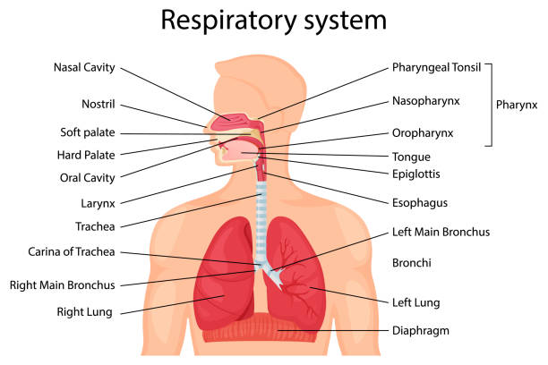
1 Q. 2 Enumerate the parts of respiratory system and write down about the lungs.
Respiratory System- The respiratory system is the set of organs that allows a person to breathe and exchange oxygen and carbon dioxide throughout the body. The integrated system of organs involved in the intake and exchange of oxygen and carbon dioxide between the body and the environment and including the nasal passages, larynx, trachea, bronchial tubes, and lungs.
The respiratory system performs two major tasks-
- Exchanging air between the body and the outside environment known as external respiration.
- Bringing oxygen to the cells and removing carbon dioxide from them referred to as internal respiration.
Parts of respiratory system
- Nose and nasal cavity.
- Pharynx
- Larynx
- Trachea
- Two bronchi
- Bronchioles
- Two Lungs
1. Nose
- Also called external nares.
- Divided into two halves by the nasal septum.
- Contains the paranasal sinuses where air is warmed.
- Contains cilia which is responsible for filtering out foreign bodies.
2. Pharynx
- Common space used by both the respiratory and digestive systems.
- Commonly called the throat.
- Originates posterior to the nasal and oral cavities and extends inferiorly near the level of the bifurcation of the larynx and esophagus.
- Common pathway for both air and food.
- Walls are lined by a mucosa and contain skeletal muscles that are primarily used for swallowing.
- Flexible lateral walls are distensible in order to force swallowed food into the esophagus.
3. Larynx
- Voice box is a short, somewhat cylindrical airway ends in the trachea.
- Prevents swallowed materials from entering the lower respiratory tract.
- Conducts air into the lower respiratory tract.
- Produces sounds.
- Supported by a framework of nine pieces of cartilage (three individual pieces and three cartilage pairs) that are held in place by ligaments and muscles.
4. Trachea
- A flexible tube also called windpipe.
- Extends through the mediastinum and lies anterior to the esophagus and inferior to the larynx.
- Cartilage rings reinforce and provide rigidity to the tracheal wall to ensure that the trachea remains open at all times.
- At the level of the sternal angle, the trachea bifurcates into two smaller tubes, called the right and left primary bronchi.
5. Two bronchi
The two primary bronchi when the trachea divides about the level of T-5.
The right bronchus
- This is wider,shorter and more vertical than the left bronchus.
- Length-2.5cm
- After entering the right lung,it divides into 3 branches,one to each lobe.
The left bronchus
- This is narrower than the right
- Length-5cm
- After entering the left lung,it divides into 2 branches,one to each lobe.
Function
- Control of air entry
- Warming & humidifying
- Support & patency
- Removal of particulate matter
- Cough reflex
6. Bronchioles
- Each lobule is supplied with air by a terminal bronchiole
- Which further subdivides into respiratory bronchioles,alveolar ducts and large numbers of alveoli (air sacs)
- About 150 million alveoli in the adult lung
- In these structures that the process of gas exchange occurs.
7. Lungs
- Each lung has a conical shape. Its wide, concave base rests upon the muscular diaphragm.
- Its superior region called the apex projects superiorly to a point that is slightly superior and posterior to the clavicle.
- Both lungs are bordered by the thoracic wall anteriorly, laterally, and posteriorly, and supported by the rib cage.
Lungs are a pair of respiratory organs situated in a thoracic cavity. Right and left lung are separated by the mediastinum.
- Texture -Spongy
Color –
- Young – brown or Grey in color.
- Adults -Mottled black due to deposition of carbon particles.
Weight-
- Right lung - 600 gms It is about 50- 100g heavier than the left lung.
- Left lung - 550 gms
Features
- Each lung is conical in shape
- It has-
- Apex at the upper end
- Base resting on the diaphragm
- Three borders,i.e.,anterior,posterior, and inferior
- Two surfaces,i.e.costal and medial
1. The apex lies above the level of first rib.It reaches 2.5 cm above the medial one third of clavicle,just medial to supraclavicular fossa.
2. The base rests on the diaphragm which separates the right lung from the right lobe of the liver and the left lung from the left lobe of the liver,fundus of stomach and the spleen
3. The anterior border of the left lung shows a wide cardiac notch below the level of the fourth costal cartilage.The heart and pericardium are uncovered by the lung in the region of this notch.
4. The posterior border corresponds to the medial margins of the heads of the ribs.It extends from the level of 7th cervical spine to the 10th thoracic spine.
5. Inferior border separates the base from costal and medial surfaces.
6. The costal surface is large and convex.It is in contact with the costal pleura and the overlying thoracic wall.
7. The medial surface is divided into a vertebral part and mediastinal part.The mediastinal part shows a cardiac impression ,the hilum.
Fissures and lobes of the lungs
The right lung is divided into three lobes by two fissures, oblique and horizontal, the left lung is divided into two lobes by oblique fissure.
- The oblique fissure cuts into whole thickness of the lung,except at the hilum
- Due to the oblique plane of the fissure ,the lower lobe is more posterior and the upper and the middle lobe more anterior
- In the right lung, the horizontal fissure passes from the anterior border upto the oblique fissure and separates a wedge shaped middle lobe from the upper lobe.
- The tongue shaped projection of the left lung below the cardiac notch is called lingula. It corresponds to middle lobe of the right lung
- The lungs expand maximally in the inferior direction because movements of the thoracic wall and diaphragm are maximal toward the base of the lung
- The presence of the the oblique fissure of the each lung allows a more uniform expansion of the whole lung.
Root of the Lung
- The root of the lung is a short ,broad pedicle which connects the medial surface of the lung to the mediastinum.
- It is formed by the structures which either enter or come out of the lung at the hilum
- The roots of lungs lie opposite the bodies of fifth,sixth and seventh thoracic vertebrae.
Contents of Root of the Lungs
- Principal bronchus on the left side, and eparterial and hyparterial bronchi on right side
- One pulmonary artery
- Two pulmonary veins, superior and inferior
- Bronchial arteries ,one on the right side and two on the left side
- Bronchial veins
- Anterior and posterior pulmonary plexuses of nerves
- Lymphatics of lung
- Bronchopulmonary lymph nodes
- Areolar tissue
Broncho Pulmonary Segment
- These are well defined sectors of the lung each one of which is aerated by a tertiary or segmental bronchus
- Each segment is pyramidal in shape with its apex directed towards the root of the lung
- These bronchopulmonary segments are independent respiratory units.


Arterial supply
- On the right side there is one bronchial artery which arises from either the third posterior intercostal artery or from the upper left bronchial artery
- On the left side there are two bronchial arteries both of which arise from the descending thoracic aorta
- There are pre capillary anastomoses between bronchial and pulmonary arteries. these connections enlarge when any one of them is obstructed in disease
Venous Drainage
- Usually there are two bronchial veins on each side ,the right bronchial vein drain into the azygous vein
- The left bronchial vein drains either into the left superior intercostal vein or into the hemi azygous vein
- The greater part of the venous blood from the lung is drained by the pulmonary veins.
Nerve Supply
- Para sympathetic nerves are derived from the vagus .
- The sympathetic nerves are derived from second to fifth spinal segments.
- These are inhibitory to the smooth muscles and glands of bronchial tree.
Q. 2. Short notes
1. Draw a labelled diagram of the eyeball.
:max_bytes(150000):strip_icc()/GettyImages-695204442-b9320f82932c49bcac765167b95f4af6.jpg)
Q .2 Structure Of Cell
If we study a cell under a microscope, we would come across three features in almost every cell-plasma membrane, nucleus and cytoplasm.
All activities inside the cell and interactions of the cell with its environment are possible due to these features.
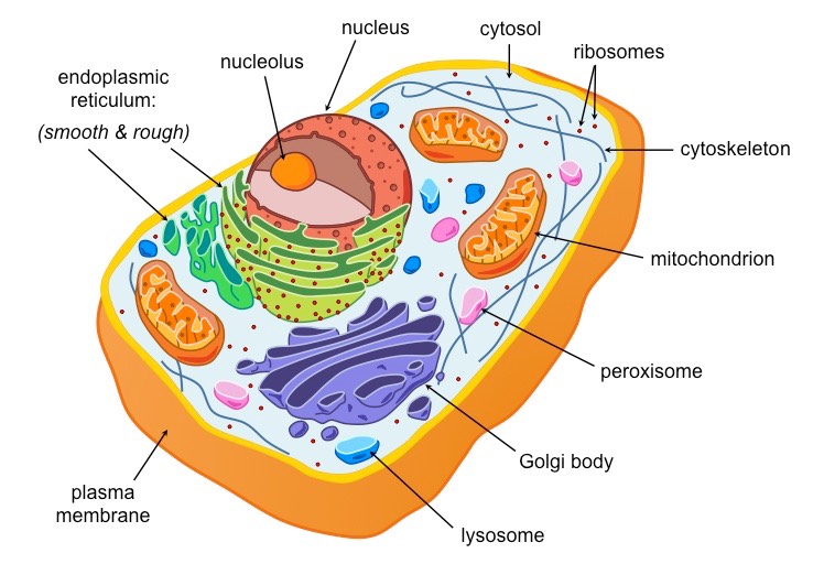
- Plasma Membrane
- Nucleus
- Cytoplasm
a. Cytosol
b. Cell Organelles
- Endoplasmic reticulum
- Golgi body
- Lysosomes
- Vacuoles
- Mitochondria
- Centrosome
- Cytoskeleton
1. Plasma Membrane
- Extremely delicate, thin , elastic, living and semi-permeable membrane
- Made up of two layers of lipid molecules in which protein molecules are floating
- Thickness varies from 75-110 A˚
- Can be observed under an electron microscope only
2. Cell wall
- Non-living and outermost covering of a cell (plants & bacteria)
- Can be tough, rigid and sometimes flexible
- Made up of cellulose, hemicellulose and pectin
- May be thin or thick, multilayered structure
- Thickness varies from 50-1000 A˚ Pectin
3. Nucleus
- Dense spherical body located near the centre of the cell
- Diameter varies from 10-25 µm
- Present in all the cells except red blood cells and sieve tube cells
- Well developed in plant and animal cells
- Undeveloped in bacteria and blue-green algae (cyanobacteria)
- Most of the cells are uninucleated (having only one nucleus)
- Few types of cells have more than one nucleus (skeletal muscle cells)
- Nucleus has a double layered covering called nuclear membrane
- Nuclear membrane has pores of diameter about 80-100 nm
- Colourless dense sap present inside the nucleus known as nucleoplasm
- Nucleoplasm contains round shaped nucleolus and network of chromatin fibres
- Fibres are composed of deoxyribonucleic acid (DNA) and protein histone
- These fibres condense to form chromosomes during cell division
- Chromosomes contain stretches of DNA called genes
- Genes transfer the hereditary information from one generation to the next.
Cytoplasm-
- Jelly-like material formed by 80 % of water
- Present between the plasma membrane and the nucleus
- Contains a clear liquid portion called cytosol and various particles Particles are proteins, carbohydrates, nucleic acids, lipids and inorganic ions
- Also contains many organelles with distinct structure and function
- Some of these organelles are visible only under an electron microscope Granular and dense in animal cells and thin in plant cells Cytoplasm Organelles
Endoplasmic Reticulum -
- Network of tubular and vesicular structures which are interconnected with one another
- Some parts are connected to the nuclear membrane, while others are connected to the cell membrane
- Two types- a. smooth(lacks ribosomes) b. rough(studded with ribosomes)
Golgi body
- Discovered by Camillo Golgi
- Formed by stacks of 5-8 membranous sacs
- Sacs are usually flattened and are called the cisternae
- Has two ends: cis face situated near the endoplasmic reticulum and trans face situated near the cell membrane
Lysosomes-
- Small, spherical, single membrane sac
- Found throughout the cytoplasm
- Filled with hydrolytic enzymes
- Occur in most animal cells and in few type of plant cells
- Help in digesting of large molecules
- Protect cell by destroying foreign invaders like bacteria and viruses
Vacuoles-
- Single membrane sac filled with liquid or sap (water, sugar and ions)
- In animal cells, vacuoles are temporary, small in size and few in number
- In plant cells, vacuoles are large and more in number
- May be contractile or non-contractile.
Mitochondria
- Small, rod shaped organelles bounded by two membranes inner and outer
- Outer membrane is smooth and encloses the contents of mitochondria
- Inner membrane is folded in the form of shelf like inward projections called cristae
- Inner cavity is filled with matrix which contains many enzymes
- Contain their own DNA which are responsible for many enzymatic actions.
- Synthesize energy rich compound ATP
Centrosome
- Centrosome is the membrane bound organelle present near the nucleus
- Consists of two structures called centrioles
- Centrioles are hollow, cylindrical structures made of microtubules Centrioles are arranged at right angles to each other
- Form spindle fibres which help in the movement of chromosomes during cell division
- Help in the formation of cilia and flagella Centrosome Centrosome matrix Centrioles Microtubules
Cytoskeleton
- Formed by microtubules and microfilaments
- Microtubules are hollow tubules made up of protein called tubulin
- Microfilaments are rod shaped thin filaments made up of protein called actin.
2. Q.3 Name any five cranial nerves and their functions.
There are 12 pairs of cranial nerves that supply structures in the head, neck, thorax and abdomen.
A cranial nerve can be made up of a mixture of functions which are called modalities or may be made up of a single modality. A modality is sensory, motor, special sensory, etc
I. Olfactory nerve
- Function: smell
II. Optic nerve
- Function: vision
III. Oculomotor
- Function: Rises upper eyelids
IV. Trochlear Nerve
- Function: Traction of eyeball laterally & downward
V Trigeminal nerve
- Function: Cornea Skin of forehead Face Mandible
VI. Abducent nerve
- Function: Lateral Rectus muscles of eyeball, turn eye laterally
VII. Facial
- Function: Muscles of face, cheek, and scalp stapedius muscle of middle ear
VIII. Vestibulocochlear
- Function: Position and movement of head Hearing
IX. Glossopharyngeal
- Function: assists swallowing
- Parotid salivary gland
X. Vagus
- Function: Constrictor muscles of pharynx and intrinsic muscles of larynx; involuntary muscle of trachea and bronchi, heart, alimentary tract from pharynx to splenic flexure of colon; liver and pancreas
XI. Accessory
- Function Muscles of soft palate, pharynx, and larynx , Sternocleidomastoid and trapezius muscles
XII. Hypoglossal
- Function: Muscles of tongue controlling its shape and movement
Q.2 4. Explain accessory organ of digestion.
Accessory” Organ-
The digestive system has several accessory organs that aid in the process of digestion. An accessory organ is one that helps with the breakdown of food without the food ever traveling through them they are not part of the digestive tract or tube.
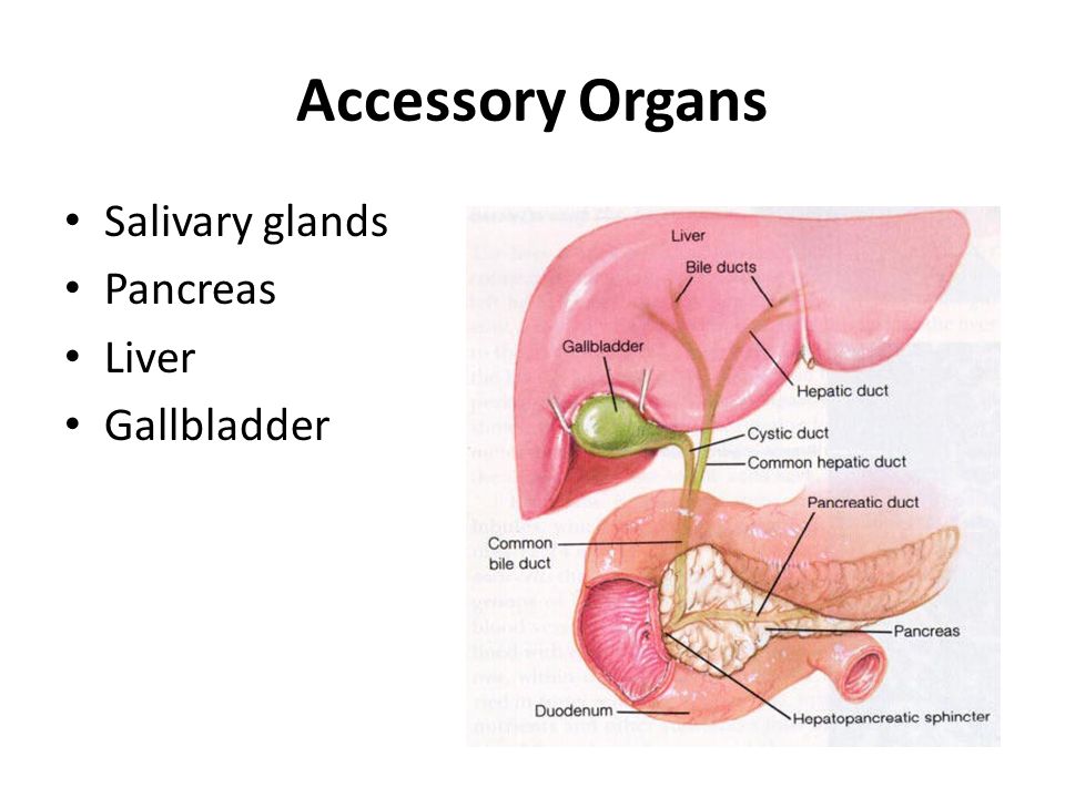
The accessory organs of digestion include-
- Teeth
- Tongue
- Salivary glands
- Exocrine part of pancreas
- Liver
- Gallbladder
Salivary glands
Major glands are
- Parotid glands
- Submaxillary or submandibular glands
- Sublingual glands
Minor salivary glands
- Lingual Mucus Glands
- Lingual Serous Glands
- Buccal Glands
- Labial Glands
Functions of saliva
- Preparation of food for swallowing
- Appreciation of taste
- Digestive function- Salivary amylase, maltase and lingual lipase.
- Cleansing and protective functions
Pancreas
- Exocrine part of pancreas resembles salivary gland in structure. It is made up of acini or alveoli.
- Each acinus has a single layer of acinar cells with a lumen in the center. Acinar cells contain zymogen granules, which possess digestive enzymes.
- In some persons, an accessory duct called duct of Santorini exists. It also opens into duodenum, proximal to the opening of ampulla of Vaterulla of vat.
Nerve supply
- Pancreas is supplied by both sympathetic and para-sympathetic fibers.
- Sympathetic fibers are supplied through splanchnic nerve.
- Para-sympathetic are supplied through vagus nerve.
Function of pancreatic juice
- Digestion of protein
- Digestion of carbohydrate
- Digestion of lipids
Liver
- Dual organ- both secretory and excretory function
- Largest gland of body
- Weight 1.5kg
- Located upper and right side of abdominal cavity
Functional anatomy of liver
- Hepatic lobes- liver is made up of many lobes which consist many hepatic lobules.
- Hepatic lobules- structural and functional unit of liver made up of hepatocyte.
- Hepatocytes -chief cells of liver.
- Hepatic plates- hepatocytes arranged in column to form plates.
- Portal triads- each portal triad consist of bile duct, hepatic artery and portal vein.
Blood supply of liver
- 1.5 l/min
- Receive blood from hepatic artery (Oxygenated) and portal vein(Deoxygenated)
- Drains blood into hepatic vein which opens into IVC
Functions of liver
- Metabolic function- carbohydrates, proteins, fats and vitamins.
- Storage function- glycogen, amino acid, iron and folic acid.
- Synthetic function- glucose (gluconeogenesis), clotting factors, steroid and heparin.
- Excretory function- cholesterol, bile salt, toxins.
- Heat production, hemopoietic and hemolytic function.
- Bile secretion
Gallbladder
- Small pear shaped organ located under the liver.
- Storage of bile
- Concentration of bile
- Alteration of pH of bile
- Secretion of mucin
- Maintenance of pressure in biliary system
- Capacity- 50ml
Q. 3. Very short questions
1. List down any four functions of growth hormone.
Ans-
- Other names- Somatotropic Hormone or Somatotropin
- Secreted by- Acidophils of Anterior Pituitary Gland
Action / Function of growth hormones
1. Growth promoting actions-
- Promotes Linear growth
- Bone – stimulate osteoblastic activity converts cartilage into bone
- Increases bone mass.
- Cartilage - stimulate proliferation of chondrocytes in epiphyseal end plate of long bones.
2. Metabolic action
- Protein metabolism
- Fat metabolism
- Carbohydrate metabolism.
- Mineral metabolism.
3. Lactogenic actions.
- Enhances milk production
- Acts like Prolactin so called Prolactin like effect of growth hormone.
Q.2 Discuss the classification of joint.
- Joint is a junction between two or more bones or cartilages. It is a device to permit movement.
- Joint are classified into structural and functional.
- Structural classification is determined by how the bones connect to each other, while functional classification is determined by the degree of movement between the articulating bones.
1. Structural classification
- Fibrous(Fixed)
- Cartilaginous(Slightly movable)
- Synovial freely(Movable)
2. Functional Classification
- Synarthrosis - Synarthroses permit little or no mobility. Most synarthrosis joints are fibrous joints.Egcranial sutures in adult.
- Amphiarthrosis -Amphiarthroses permit slight mobility. The two bone surfaces at the joint are both covered in hyaline cartilage and joined by strands of fibrocartilage. eg: secondary cartilaginous joints
- Diarthrosis- Permit a variety of movements. Only synovial joints are diarthrodial.
3. Classification According to number of articulating bones
- Simple Joint- 2 articulation surfaces (eg. shoulder joint, hip joint)
- Compound Joint- 3 or more articulation surfaces (eg. radiocarpal joint)
- Complex Joint- 3 or more articulation surfaces and an articular disc or meniscus (eg . knee joint)
Q. 3 Name three middle ear bones.
Ans-The Auditory Ossicles/Ear Bones are-
- Malleus- It is the outermost of the three bones, shaped like a hammer, hence the name (malleus is the Italian for hammer).
- Incus- The incus or anvil is an anvil-shaped bone located after the malleus, consisting of a body and long and short limbs.
- Stapes- The smallest of the three bones, it is located in the innermost part of the middle ear. This stirrup-shaped bone has four parts The head, base, and the anterior and posterior limbs. posterior limbs.
Q.4 What is meant by voluntary muscles?
- A voluntary muscle is a muscle that you choose to move, like those in the arms and legs, as opposed to the ones that move automatically, like the heart.
- Muscle is the tissue in animals that produces movement or motion. Voluntary means done out of free will or by choice.
- Voluntary muscles are also often called skeletal muscles (because all of the muscles attached to the skeleton are voluntary muscles) or striated muscles (because the muscle fibers make them look striated, or stripy).
Q.5 What is tissue? Enlist the types of tissues
Ans-
- Tissue -Is a group of similar cells specialized for the performan[1]ce of a common function.
- In biology, tissue is an assembly of similar cells and their extracellular matrix from the same embryonic origin that together carry out a specific function.
- The study of tissues is called Histology
There are 4 major groups of somatic tissues, and these are:
- Epithelial Tissues
- Connective Tissues
- Muscular Tissues
- Nervous Tissues
Section-B (Applied Physiology)
Q1.1 Describe the process of oxygen transport from lungs to the tissues.
- Blood serves to transport the respiratory gases.
- Oxygen, which is essential for the cells is transported from alveoli of lungs to the cells.
- Carbon dioxide, which is the waste product in cells is transported from cells to lungs.
Transport of Oyygen
Oxygen is transported from alveoli to the tissue by blood in two forms:
- As simple physical solution (3%).
- In combination with hemoglobin (97%).
1- Simple Physical Solution-
- Oxygen dissolves in water of plasma.
- 0.3 mL/100 mL • About 3% of total oxygen in blood.
- Poor solubility of oxygen in water content of plasma.
- During the exercise this type of oxy transport is used to meet the excess demand of oxygen by the tissues.
2-In Combination With Hemoglobin[-
- Oxygen combines with hemoglobin in blood.
- Transported as oxyhemoglobin.
- Maximum amount (97%).

Oxygenation of Hemoglobin
- Oxygen combines with hemoglobin.
- In the process of oxygenation and not oxidation (does not change the ferrous of Hemoglobin).
- Oxygen can be readily released from hemoglobin when it is needed.
Oxygen Carrying Capacity of Hemoglobin-
- Oxygen carrying capacity of hemoglobin is the amount of oxygen transported by 1 gram of hemoglobin.
- It is 1.34 mL/g.
Oxygen Carrying Capacity of Blood-
- Oxygen carrying capacity of blood refers to the amount of oxygen transported by blood.
- Normal hemoglobin content in blood is 15 g%.
- Since oxygen carrying capacity of hemoglobin is 1.34 mL/g, blood with 15 g% of hemoglobin should carry: 1.34 X 15 = 20.1 mL% of oxygen, i.e. 20.1 mL of oxygen in 100 mL of blood.
Remember-
- Each Hb can carry 4 oxygen.
- One RBC contains 250 million Hb molecules, so can carry 1 billion oxygen.
- 1g of Hb combine 1.34ml of Oxygen.
- 100ml of blood contains 15g of Hb, so 15g X 1.34ml/g = 20 ml of oxygen.
Saturation of Hemoglobin with Oxygen-
- Saturation is the state or condition when hemoglobin is unable to hold or carry any more oxygen.
- Saturation of hemoglobin with oxygen depends upon partial pressure of oxygen (a rise in PaO2 is accompanied by a rise in the arterial oxygen saturation).
- And it is explained by oxygen- hemoglobin dissociation curve.
The number of occupied O2- binding sites on the Hb molecule:
- 100% = 4 binding O2. – PO2 = 95 mm Hg
- 75% = 3 binding O2. – PO2 = 40 mmHg
- 50% = 2 binding O2. – PO2 = 25 -27 mmHg
- 25% = 1 binding O2. – PO2 = 15 mmHg
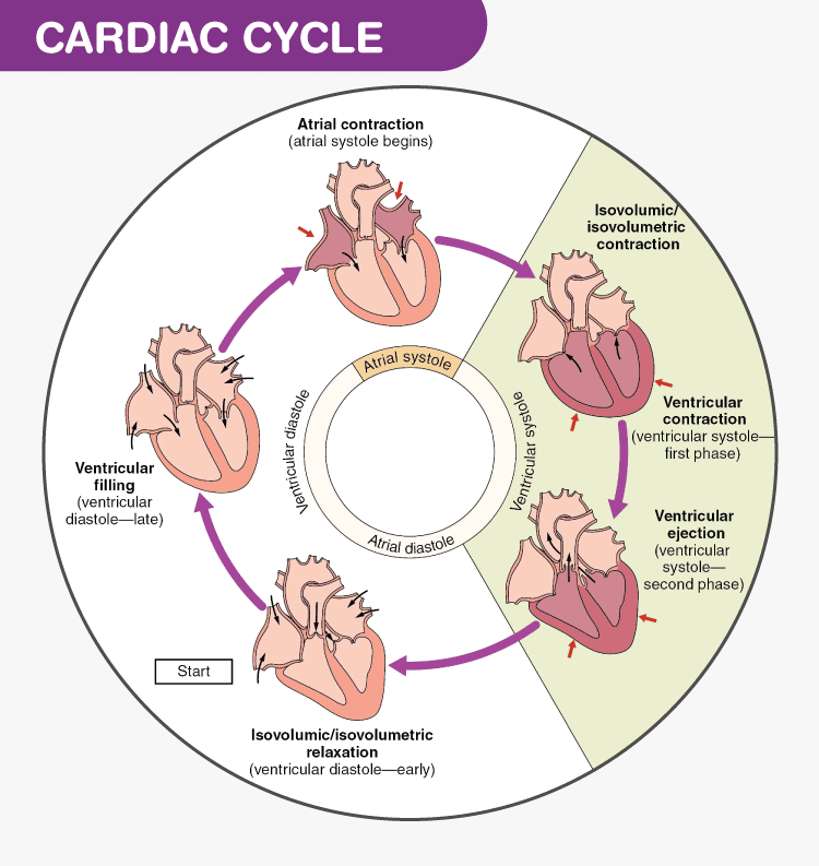
Q1. 2 Define and draw a diagram of cardiac cycle. Describe the ventricular events in cardiac cycle.
Cardiac Cycle
Definition-
- The sequence of changes in the pressure and flow in the heart chambers and blood vessels in between the two subsequent cardiac contractions is known as cardiac cycle.
- Normal duration: 0.8 second at heart rate of 75 per minute.
- Atrial systole: 0.1 second
- Ventricular systole: 0.3 second
- Atrial diastole: 0.7 second
- Ventricular diastole: 0.5 second
Events in Cardiac Cycle-
- The parts of heart normally beat in an orderly sequence- atrial systole, ventricular systole, atrial diastole and ventricular diastole.

1. Atrial Systole
- Duration 0.1 second. It is seen following the impulse generation in the SAN.
- Atrial muscle contracts and atrial pressure rises with ventricular pressure following it.
- It propels approximately 30% additional blood into the ventricles.
2. Ventricular Systole
After excitation of the atria, the impulse from the SA node pass to AV node by internodal fibres and further down to the ventricular muscle via the His Purkenji system and cause ventricular depolarization of ventricular muscle. Considering a cardiac cycle time of 0.8 sec, the duration of ventricular systole is 0.3 sec and it has three phases.
a. Isovolumetric Contraction
With the onset of ventricular systole, the pressure within the ventricle rises and to prevent backflow of blood from ventricles to the atria, the AV valves close. This produces the first heart sound. This time the semilunar vales remain closed. So the ventricles contract as a closed chamber with much rise in intraventricular pressure, but without any significant rise in volume. So, this is called isovolumetric contraction phase.
b. Rapid Ejection Phase
As the name implies, in this phase, blood is ejected rapidly from ventricles into the arteries. After isovolumetric phase, ventricular contraction continues, there is further rise in intraventricular pressure. When the pressure becomes more than the aorta and pulmonary artery, the semilunar valves open and blood is ejected forcefully into the aorta (from left ventricle) and pulmonary artery (from right ventricle). Due to forceful ejection, this is called rapid ejection phase.
c. Slow Ejection Phase
With rapid ejection, as much of the blood volume is ejected out, the intraventricular pressure falls, and now, blood continues to be ejected slowly for a longer period. This phase is therefore called slow ejection phase. However, the volume of blood ejected in this phase is less than the volume ejected in rapid ejection phase.
3. Atrial Diastole-
- Duration 0.7 second.
- During this phase, atrial muscles relax and atrial pressure gradually increases due to continuous venous return.
4. Ventricular Diastole-
Given a cardiac cycle time of 0.8 sec, the ventricular diastolic time is 0.5 sec and it comprised of five different phases, each marked with different events. These phases are as follow:
a. Protodiastole
This is the very brief initial phase of ventricular diastole. As the ventricles start relaxing, the intraventricular pressure declines and blood in the aorta and pulmonary arteries moves back towards the ventricles. This closes the semilunar valves and the second heart sound is produced.
b. Isometric Relaxation Phase
This begins with the closure of semilunar valves. As the AV valves are already closed, the ventricles relax as a closed chamber with rapid decline in pressure with volume remaining same, so this phase is termed as isovolumetric relaxation.
c. 1st Rapid Filling Phase
During ventricular diastole, the atria also remain in diastolic phase. The right and left atrium continue to be filled with blood returning via the vena cava and pulmonary veins respectively, which raises the intra atrial pressure. However, at the same time, the intraventricular pressure is reduced due to further ventricular relaxation. When the atrial pressure become more than the ventricular pressure, the AV valves open which allows a rapid initial flow of blood into the ventricles. During this time the ventricular relaxation continues and both intratribal and intraventricular pressure falls. The initial rapid flow of blood from atria to the ventricles produces the 3rd heart sound.
d. Slow Filling or Diastasis
In this phase, as the intra atrial pressure becomes very less, the force generated by the atria to move blood into the ventricles is also reduced. Blood moves very slowly and passively from atria to the ventricles and this is termed slow filling phase or diastasis. Almost 75% of ventricular filling occurs during rapid and slow filling phase.
e. Last rapid Filling Phase
This is the last phase of ventricular diastole that coincides with atrial systole. As atrial systole begins, pressure within the atria is raised and blood is once again forced into the ventricles, which marks the last rapid filling.
2. Short notes (Attempt any three): 3x5=15
Q.1 Enumerate the different blood groups and the importance of determination of blood groups.
- Dr. Karl Landsteiner's discovered the ABO Blood Group System in 1901.
- He and five co-workers began mixing each others red blood cells and serum together and accidentally performed the first forward and reverse ABO groupings.
- Blood group, also known as blood type, is a category (type) of blood groups based on the presence and absence of blood group antigens in the surface (membrane) of the RBCs, and antibodies on the plasma.
Antigen
- An antigen is a substance usually a protein which when introduced into an individual who recognizes it as foreign, leads to the production of antibody. This antibody specifically reacts with the antigen. On the red cell surface there is presence of glycoproteins and glycolipids which act as antigens. They are called blood group antigens. These antigens can be on the surface, below or protrude from the red cell membrane. If introduced into the body of an individual who lacks the antigen, an immune reaction can occur.
Antibodies
- These are immunoglobulins present in the serum and can be of 5 types: IgG, IgM, IgD, IgA and IgE.
- If red cells carrying an antigen are introduced into the circulation of an individual who lacks that antigen, antibodies will form and cause destruction of the introduced red cells.
- These are immune or acquired antibodies and are IgG in nature.
- They react best at 37°C.Certain antibodies occur without antigenic stimulus and are called naturally occurring antibodies e.g. ABO antibodies. They are IgM in nature and reactat room temperature.
The ABO grouping system
The ABO grouping system is subdivided into 4 types based on the presence or absence of antigens A and B on the red cell surface as shown below.
- Red cells that only have antigen A are called group A.
- Those that only have B antigen are called group B.
- Cells that have both A and B antigens are group AB.
- Cells that lack both antigens are O
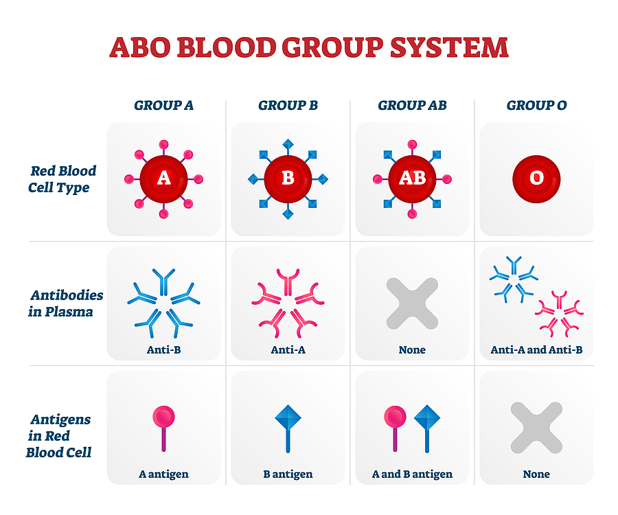
- Blood having antigen A belongs to A group. This blood has B antibody in the serum.
- Blood with antigen B and a antibody belongs to B group.
- If both the antigens are present, blood group is called AB group and serum of this group does not contain any antibody.
- If both the antigens are absent, blood group is called O group and serum of this group contain any a & b antibodies are present in the serum.
- Thus, RBC of "O" group has no antigen and so agglutination does not occur with any other group of blood. So,"O" group blood can be given to any blood group persons and the people with this blood group are called universal donors
- Plasma of AB group blood has no antibody. This does not cause agglutination of RBC from any other group of blood. People with AB group can receive blood from any blood group persons. So, people with this blood group are called universal recipients.
Importance of determination of blood groups.
- The red cells of an individual contain antigens on their surfaces that correspond to their blood group and antibodies in the serum that identify and combine with the antigen sites on the surfaces of red cells of another type.
- The reaction between red cells and corresponding antibodies usually results in clumping agglutination of the red cells therefore, antigens on the surfaces of these red cells are often referred to as agglutinogens.
- Antibodies are part of the circulating plasma proteins known as immunoglobulins, which are classified by molecular size and weight and by several other biochemical properties.
- Most blood group antibodies are found either on immunoglobulin G (IgG) or immunoglobulin M (IgM) molecules, but occasionally the immunoglobulin A (IgA) class may exhibit blood group specificity. Naturally occurring antibodies are the result of immunization by substances in nature that have structures similar to human blood groups.
- These antibodies are present in an individual despite the fact that there has been no previous exposure to the corresponding red cell antigens for example, anti-A in the plasma of people of blood group B and anti-B in the plasma of people of blood group A. Immune antibodies are evoked by exposure to the corresponding red cell antigen. Immunization (i.e., the production of antibodies in response to antigen) against blood group antigens in humans can occur as a result of pregnancy, blood transfusion, or deliberate immunization.
- The blood group antigens are not restricted solely to red cells or even to hematopoietic tissues. The antigens of the ABO system are widely distributed throughout the tissues and have been unequivocally identified on platelets and white cells (both lymphocytes and polymorphonuclear leukocytes) and in skin, the epithelial (lining) cells of the gastrointestinal tract, the kidney, the urinary tract, and the lining of the blood vessels.
- Evidence for the presence of the antigens of other blood group systems on cells other than red cells is less well substantiated. Among the red cell antigens, only those of the ABO system are regarded as tissue antigens and therefore need to be considered in organ transplantation.
Q.2 Functions of thyroid hormone
Functions Of Thyroid Hormones
- Action On Basal Metabolic Rate (BMR) -Increases the metabolic activities in most of the body tissues, except brain, retina, spleen, testes and lungs. It increases BMR by increasing the oxygen consumption of the tissues.
- Action On Protein Metabolism-Increases the synthesis of proteins in the cells.
- Action On Carbohydrate Metabolism-Stimulates almost all processes involved in the metabolism of carbohydrate.
- Action On Fat Metabolism -Decreases the fat storage by mobilizing it from adipose tissues and fat depots.The mobilized fat is converted into free fatty acid and transported by blood.
- Action On Plasma And Liver Fats- Even though there is an increase in the blood level of free fatty acids, thyroxine specifically decreases the cholesterol, phospholipids and triglyceride levels in plasma.
- Action On Vitamin Metabolism- Thyroxine increases the formation of many enzymes.Since vitamins form essential parts of the enzymes, it is believed that the vitamins may be utilized during the formation of the enzymes.
- Action On Body Temperature- Thyroid hormone increases the heat production in the body, by accelerating various cellular metabolic processes and increasing BMR. It is called thyroid hormone induced thermogenesis.
- Action On Growth -Thyroxin has both general and specific effects on growth. Increase in thyroxine secretion accelerates the growth of the body, especially in growing children.
- Action On Body Weight- Thyroxine is essential for maintaining the body weight. Increase in thyroxine secretion decreases the body weight and fat storage. Decrease in thyroxine secretion increases the body weight because of fat deposition.
- Action On Blood- Thyroxine accelerates erythropoietic activity and increases blood volume. It is one of the important general factors necessary for erythropoiesis.
- Action On Cardiovascular System-Thyroxine acts directly on heart and increases the heart rate.
- Action On Respiration-Thyroxine increases the rate and force of respiration indirectly.
- Action On Gastrointestinal Tract-Generally, thyroxine increases the appetite and food intake. It also increases the secretions and movements of GI tract.
- Action On Central Nervous System- Thyroxine is very essential for development of CNS Very important to promote growth and development of the brain during fetal life and during the first few years of postnatal life.Thyroid deficiency in infants results in abnormal development of synapses, defective myelination and mental retardation.
- Action On Skeletal Muscle-Thyroxine is essential for the normal activity of skeletal muscles. Slight increase in thyroxine level makes the muscles to work with more vigor.
- Action On Sleep-Normal thyroxine level is necessary to maintain normal sleep pattern.Hypersecretion of thyroxine causes excessive stimulation of the muscles and central nervous system.
- Action On Sexual Function -Normal thyroxine level is essential for normal sexual function. In men, hypothyroidism leads to complete loss of libido (sexual drive) and hyperthyroidism leads to impotence.
- Action On Other Endocrine Glands-Because of its metabolic effects, thyroxine increases the demand for secretion by other endocrine glands.
Q.2- 3.Functions of white blood cells.
White blood cells, also known as leukocytes, are responsible for protecting your body from infection. As part of your immune system, white blood cells circulate in your blood and respond to injury or illness.
Leucopoiesis- The process of development and maturation of white blood cells(leucocytes), is called leucopoiesis.
Functions
1. Neutrophils
- 1st line of defense
- Granules contain enzymes like Nucleotidases Catalases
- Antibody like substances -> Defensins
2. Eosinophils
- Defense (specially against parasites )
- Role in allergic reactions
- Substances present in granules 1. Major basic protein 2. Eosinophilic cationic proteins 3. Eosinophil peroxidase
3. Basophils
- Role in allergic responses
- Substances present in granules 1. Histamine 2. Heparin 3. Hyaluronic acid 4. Proteases & Myeloperoxidase
4. Monocytes
- 1st line of defense
- Motile & phagocytic
- Precursors of tissue macrophages secrete: 1. Interleukin 1 2. Colony stimulating factor 3. Platelet activating factor
5. Lymphocytes
- Immunity
- T- Lymphocyte -> Cellular Immunity
- B- Lymphocyte -> Humoral Immunity
Q.2 .4 Explain the mechanism of muscle contraction
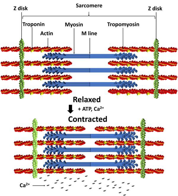
Ans- Contraction -The Slide Filament Mechanism
All parts of the muscle fiber and the electrical changes mentioned above are involved in the process of contraction, which is the gliding thread mechanism. Nerve impulses cause muscle fiber depolarization and this electrical change allows the myosin filaments to pull the actin filaments toward the center of the sarcomere, shortening the sarcomere. All sarcomere shortens and muscle fibers contract. A more detailed description of this process is as follows-
- Nerve impulses reach axon terminals. Acetylcholine is released and diffuses throughout the synapse.
- Acetylcholine increases the permeability of the sarcolemma to Na+ ions entering the cell.
- The sarcolemma becomes depolarized, negative on the outside and positive on the inside. T-tubules carry the reversal of charges to the interior of the muscle cell.
- Depolarization stimulates the release of Ca+2 ions from the sarcoplasmic reticulum. Ca+2 ions bind to the troponin-tropomyosin complex and leave the actin filament.
- Myosin breaks down ATP to release energy. Bridges on myosin attach to actin filaments and pull them toward the center of the sarcomere, shortening the sarcomere.
- All the sarcomere shortening of the muscle fiber the entire muscle fiber contracts. (Meaning not clear)
- The sarcolemma repolarizes. The K+ ions leave the cell and return to a positive charge on the outside and a negative charge on the inside. The pump returns Na+ ions to the outside and K+ ions to the inside.
- Sarcolemma cholinesterase inactivates acetylcholine.
- A subsequent nerve impulse prolongs the contraction (more acetylcholine is released).
- Without further stimulation, the muscle fibers relax and return to their original length.
Steps 1 to 8 of this sequence represent the contraction of a single muscle fiber (called a twitch) in response to a single nerve impulse. All this takes less than a second, so if the muscle fibers relax immediately after contraction, they will not be able to move meaningfully. Normally, however, nerve impulses occur in a steady stream, causing sustained contractions called tetanus. For muscles like the biceps brachii that flex the forearm, effective movement means many of the thousands of muscle fibers are in tetanus, a state of sustained contraction. As expected for such a complex process, muscle contraction can be compromised in a number of ways.
Q 3. Very short questions (Attempt any three)-
Q.1 List down any four functions of growth hormone.
The growth hormone is produced by the anterior pituitary. It is made up of 191 amino acids that make a long single-chain polypeptide. It is synthesized in somatotropic cells found in the anterior pituitary gland.
Growth Hormone Function
Following are the important growth hormone function-
- It maintains normal body structure and metabolism.
- Maintains, builds, and repairs healthy tissue in the brain and other organs.
- The growth hormone is utilized widely in medicines that heal the growth disorders in children and hormone deficiency in adults.
- The growth hormone enhances growth in adolescents and children.
- It also contributes to the regulation of body fluids, fat metabolism, sugar and also the functions of the heart.
- The growth hormone reduces body fat by increasing bone density and muscle mass.
- The energy levels rise consequently, along with improved skin tone and bone density.
Q.3- 2. What is vital capacity and its normal value?
Vital capacity-Vital capacity (VC) is the maximum amount of air a person can expel from the lungs after a maximum inhalation. It is equal to the sum of inspiratory reserve volume, tidal volume, and expiratory reserve volume.
- Vital capacity is the amount of air that can be exhaled after a maximal inhalation.
- It depends on factors such as age, sex, height, mass, and possibly ethnicity.
- A normal adult has a vital capacity between 3 and 5 liters
Q. 3-3 Any two functions of cell membrane.
Ans-The cell membrane is also known as the plasma membrane. It is the outermost covering of animal cells. It is a semi-permeable membrane composed of lipids and proteins. The plasma membrane forms the boundary between the outer environment and living systems.
The main functions of the plasma membrane or cell membrane include-
- Protecting the integrity of the interior cell.
- Providing support and maintaining the shape of the cell.
- Helps in regulating cell growth through the balance of endocytosis and exocytosis.
- The cell membrane also plays an important role in cell signalling and communication.
- It acts as a selectively permeable membrane by allowing the entry of only selected substances into the cell.
Q.3- 4 List any two physiological properties of skeletal muscles.
Ans-Properties Of Skeletal Muscle
The skeletal muscles have the following properties-
- Extensibility- It is the ability of the muscles to extend when it is stretched.
- Elasticity- It is the ability of the muscles to return to its original structure when released.
- Excitability- It is the ability of the muscle to respond to a stimulus.
- Contractility- It is the ability of a muscle to contract when in contact with a stimulus.