Bacteria
Bacteria-
Bacterial are unicellular prokaryotic, microscopic organisms and the unit of measurement used for measuring bacteria is micron (1 micron or micrometre is one thousandth of a millimetre). They vary greatly in size. The size ranges from 0.1 micron to 2 microns in width and about 3 microns to 10 microns in length but filamentous forms may even exceed 100 microns.
Bacteria are of various shapes- These may be spherical, rod shaped, straight, slightly curved (comma shaped), branched, filamentous and I, V, L shaped (arranged like Chinese letters). Sometimes the length of the organism approximates the width of the organism.
Anatomy-
The following six structures can be commonly seen in bacterial cell proceeding from outside inwards.

Flagella-
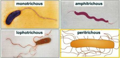
They are the organs of locomotion. It is 15-20nm hair like helical structure emerges from cell wall. They are fine hair-like protein fibrils attached to the cell in various locations and each flagellum originates from a basal granule. They are present in motile bacteria and in some spiral forms. The number and arrangement of flagella are characteristic for each bacterium.
- Monotrichate- A single flagellum at one end of the cell. Example- Cholera vibrios.
- Amphitrichate- Two flagella, one at each pole. Example- Alcaligenes faecales.
- Lophotrichate- A tuft of flagella at one or both ends. Example- Pseudomonas.
- Petritrichate-Several flagella present all over the surface. Example- Salmonella typhi.
Capsule-
Capsule is 0.2µm or more, visable under light microscope. Some bacterial cells are surrounded by a thick protective gelatinous layer known as capsule. It is concerned with the virulence of the bacterium, e.g. diplococcus pneumoniae Distinction between the cell wall and capsule is not always clear. When the capsule is in the form of a loose secretion, it is called a slime layer and if it is too thin it is known as microcapsule.
Microcapsule-Thickness less than 0.2µm, visible under Electron microscope.
Macro-Capsule- Thickness of 0.2µm or more, visible under light microscope.
Cell Wall-
It is present just beneath the capsule or slime layer. It is tough, somewhat rigid and gives shape to the cell. It forms a rigid frame and protects the cell from damage. It varies in thickness, rigidity and in chemical composition. Gram positive bacteria have a simpler chemical composition as compared to Gram negative bacteria. Bacterial cell wall is composed of cellulose, protein and lipid.
Cytoplasmic Membrane-
The cell membrane in bacteria is very thin, weak, highly flexible and it is commonly called as cytoplasmic membrane. It is a thin layer lining the inner surface of the cell wall and separates it from the cytoplasm. It acts as a semipermeable membrane and controls the passage of nutrients and waste products into and out of the cell. Damage to this membrane results in leaking the vital material out of the cell and the cell ultimately dies. Chemically it consists of lipoprotein with a small amount of carbohydrate.
Cytoplasm-
Inside the cell bounded by the cytoplasmic membrane is a clear watery substance called cytoplasm, which is slightly viscous and homogeneous in appearance. It is a colloidal system containing a variety of macromolecules (proteins, nucleic acid, polysaccharides and lipids), organic and inorganic solutes. Cytoplasm contains ribosomes, mesosomes, vacuoles and stored food granules viz. glycogen and volutin.
- Ribosomes- These are in the form of tiny granules and are the centre of protein synthesis. ribosome is of 70S type.
- Mesosomes- These are formed by the invagination of the cytoplasmic membrane and are the principal sites of the respiratory enzymes.
- Vacuoles- These are fluid containing cavities.
- Glycogen and Volutin Granules- These are stored food granules.
Nuclear Body (Nucleus)
The nucleus of bacteria for a long time was a controversial structure. In the past, some considered the entire bacterial cell as a nucleus and others regarded the staining body in the centre as nucleus.
Feulgen stain reveals the presence of primitive form of nucleus in bacteria consisting of a long filament of deoxyribose nucleic acid (DNA) tightly coiled inside the cytoplasm. It represents the chromosome and is responsible for transmission of hereditary characters.
DNA replicates and is equally distributed among the daughter cells. A bacterial cell has no nuclear membrane or nucleous. An organised nucleus as found in animals and higher plants is absent in bacteria.
Function-
- It contains and stores hereditary information of the cell.
- It controls all cell activities.
Classification of Bacteria-
The basis of their shapes they are classified into three groups which are; cocci, bacilli and spirilli
Cocci (Spherical Bacteria)-
These are spherical or oval in shape about 1 micron in diametre. Many bacteria of this form exhibit patterns of arrangement that are important for identification purposes and are characteristic of particular species of bacteria.
- Micrococci-These are found singly or in small clusters like bunches of grapes, e.g. micrococcus pyogenes causing boils on skin.
- Diplococci- These are cocci in pairs, e.g. diplococcus pneumoniae which causes pneumonia.
- Neisseria-These are cocci in pairs but bean shaped, e.g. neisseria gonorhoeae and N. meningitis.
- Streptococci-These are cocci in chains, e.g. streptococcus lactis which causes souring milk and cheese.
- Staphylococci- These are cocci in clusters, e.g. staphycoccus aureus causing pyogenic infection.
- Sarcina- These are cocci in packet arrangement with 8 or more cells. They divide in three planes at right angles to one another resulting in a regular pattern of cubes or packets of cells, e.g. sarcina lutea found in water, air and soil.
Bacilli (Rod Shaped Bacteria)-
The bacteria are cylindrical or rod shaped. Generally, they occur as single unattached cells (microbacilli) and do not arrange themselves in the variety of patterns characteristic of cocci. Occasionally, they occur in pairs (diplobacili), chains (streptobacilli), Chinese letter pattern or in the form of branched filaments.
- Microbacilli-They occur as single unattached cells, e.g. Brucella.
- Diplobacilli- They occur in pairs, e.g. klebsiella pneumoniae.
- Streptobacilli- They are found in chains, e.g. bacillus anthracis.
- Chinese letter pattern- They are arranged in straight, V, L, I, Y shapes, i.e. Chinese letter pattern, e.g. corynebacterium diphtheriae.
- Branched Filaments- They may be arranged in the form of branched filaments, e.g. actinomycetes.
- Coccobacilli- Sometimes in bacilli the length may approximate the width of the organism. Such organisms are called cocobacilli, e.g. brucella.
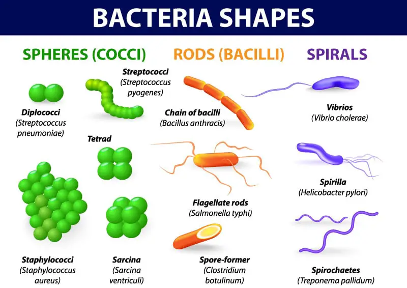
Spirilli (Spiral Shaped Bacteria)-
They are curved or spiral shaped motile bacteria. They occur mostly as unattached individuals found freely in water. The individual cells of different species show striking differences in length, number of coils and rigidity of cell walls.
- Vibrio- Vibrio is a short curved comma shaped Gram negative motile bacteria. The pathogenic species is Vibrio cholerae which causes cholera in human being.
- Spirilla- They are short rigid spiral shaped organisms found freely in water. They have very few wavy bands along the long axis, e.g. spirilla.
- Spirochactes-They are thin long spiral flexible organisms. They are able to wriggle, flex and bend themselves like coiled steel spring. Many spirochaetes are free living found in water, soil and decaying organic matters while others occur as commensals in the human mouth and genitalia and some are pathogenic to human being. One of the most important pathogenic spirochaete is Treponema pallidum which causes the veneral disease syphilis in human being.